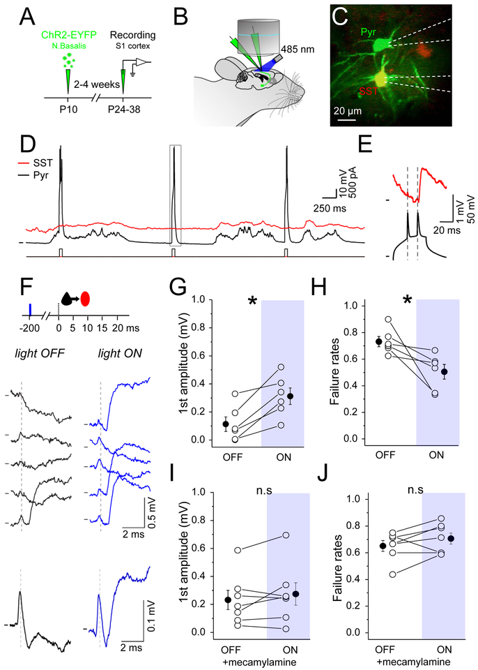
Figure 6. Nicotinic receptors enhance EPSP efficacy at Pyr-SST connections in vivo.
(A) Schematic of the experimental procedure. P10 SST-IRES-Cre-Ai9 pups were injected in nucleus basalis (Bregma: 0.02 mm, lat: 1.3 mm, depth: 4.5 mm). After an incubation period of 2–4 weeks mice were anesthetized and electrophysiological recordings were performed in somatosensory cortex. (B) Schematic of the recording setup. (C) In vivo 2-photon image of a pyramidal neuron (green cell soma) connected to a SST interneuron (yellow cell soma). White dashed lines show recording electrodes outlines. (D) Example recording of a Pyr (black trace) connected to SST (red trace). Bottom trace shows injected current into Pyr to drive spiking. (E) Zoom of the grey rectangle in (D). In this example two spikes were evoked and only the second one led to an EPSP in the SST interneuron (red trace). (F) Top: schematic of the experimental protocol: 1 blue light pulse (10 ms) was delivered on the brain surface (5-15 mm) 200 ms prior to the presynaptic Pyr doublet of spikes. Middle: example single traces showing the SST response to the first evoked Pyr spike under baseline/light OFF (black traces) and light ON (blue traces) conditions. Bottom: average responses from the above Pyr to SST connection (Light OFF, n=27 trials; Light ON, n=30 trials). (G) Mean EPSP amplitude in response to the first spike of the doublet, for both conditions. (H) Mean failure rate after the first spike, for both conditions. (I) and (J) The same as (G) and (H) but in the presence of the nicotinic receptors antagonist (mecamylamine). See also Figure S3 and S4.

