

Abstract
Climate change has accentuated the importance of understanding how organisms respond to stresses imposed by changes to their environment, like water availability. Unusual organisms, called anhydrobiotes, can survive loss of almost all intracellular water. Desiccation tolerance of anhydrobiotes provides an unusual window to study the stresses and stress response imposed by water loss. Because of the myriad of stresses that could be induced by water loss, desiccation tolerance seemed likely to require many established stress effectors. The sugar trehalose and hydrophilins (small intrinsically disordered proteins) had also been proposed as stress effectors against desiccation because they were found in nearly all anhydrobiotes, and could mitigate desiccation-induced damage to model proteins and membranes in vitro. Here, we summarize in vivo studies of desiccation tolerance in worms, yeast, and tardigrades. These studies demonstrate the remarkable potency of trehalose and a subset of hydrophilins as the major stress effectors of desiccation tolerance. They act, at least in part, by limiting in vivo protein aggregation and loss of membrane integrity. The apparent specialization of individual hydrophilins for desiccation tolerance suggests that other hydrophilins may have distinct roles in mitigating additional cellular stresses, thereby defining a potentially new functionally diverse set of stress effectors.
INTRODUCTION
For centuries, biologists have been drawn to the extreme traits of unusual organisms, such as the incredibly rapid cell division of bacteria, the huge size of amphibian nuclei or squid axons, and the extreme chromosome metabolism of ciliates. The study of extreme traits like these has led to many seminal discoveries and transformative technical advances in biology. As one example, the ciliate Tetrahymena thermophila shatters a duplicated set of its chromosomes, generating huge numbers of chromosome ends. The abundance of these ends made possible biochemical and genetic approaches that led to the discoveries of the molecular composition, function, and assembly of telomeres (the specialized structures at the end of all eukaryotic chromosomes; Blackburn et al., 2006). By analogy, can the study of rare organisms that survive extreme changes in their environment provide new insights into stress biology?
Understanding how organisms adjust to changes in their ever-changing environments has been a holy grail of biology. To complete this quest, scientists have studied different organisms after they have been stressed by typical environmental changes (e.g., small changes in temperature, food availability, or osmolarity). Through this approach, they have achieved a detailed molecular understanding of the damages that are induced by different stresses and the cellular factors (stress effectors) that either prevent or mitigate them. However, rare organisms can survive much more extreme environmental stresses. For example, centuries ago, organisms, called anhydrobiotes, were discovered that survive environmentally driven huge losses of (>95%) intracellular water. Anhydrobiotes include seeds, a subset of microbes, and some mature plants and animals (Crowe et al., 1992; Potts, 2001). The desiccation tolerance of these remarkable organisms raises many questions. What are the major stresses of desiccation? Which stress effectors are necessary for desiccation tolerance in anhydrobiotes, and how do any of these factors prevent or mitigate the damage of desiccation?
Answering these questions had been foiled by two complications. First, desiccation is likely to induce many stresses because water is a critical solvent and cofactor for so many biological processes. Therefore, which of the stress(es) contribute to the lethality of desiccation was unclear. Second, desiccation tolerance in anhydrobiotes appears only after environmental changes like starvation that lead to extensive changes in the levels and activities of many well-studied cellular proteins, including known stress effectors (Calahan et al., 2011; Erkut et al., 2011; Boothby et al., 2017). These specialized states also have elevated levels of surprising constituents including disaccharides (most commonly trehalose but occasionally others), small metabolites (e.g., proline), and a number of small intrinsically disordered proteins (Crowe et al., 1992; Potts, 2001). These disordered proteins exhibit very little sequence identity to each other both within and between species, and therefore have been grouped and named based on shared general characteristics (e.g., charge) and organism of origin (Crowe et al., 1992; Crowe, 2015; Potts, 2001; Erkut et al., 2013). For simplicity, we will refer to them by the single name, hydrophilins. Whether any of the hydrophilins or other changing cellular constituents were important stress effectors for desiccation tolerance was also unclear (Larsen, 1993; De Virgilio, 2012; Crowe, 2015). These two challenges are beginning to be overcome through studies in the nematode Caenorhabditis elegans, the budding yeast Saccharomyces cerevisiae, and the tardigrade Hypsibius exemplaris (Figure 1). The emerging results provide exciting new molecular insights into desiccation-induced stresses and desiccation tolerance. They also underscore the potential of small molecules and proteins as novel and potent stress effectors.
FIGURE 1:
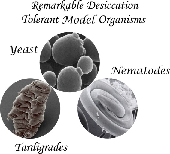
Desiccation tolerant model organisms. Recent studies in desiccation tolerant model organisms including budding yeast (Saccharomyces cerevisiae), nematodes (Caenorhabditis elegans), and tardigrades (Hypsibius exemplaris) provide new molecular insights into this rare trait.
IDENTIFYING THE STRESS EFECTORS OF DESICCATION TOLERANCE
In nematodes, desiccation tolerance is achieved when dense populations are starved to induce dauer larvae, followed by their preconditioning with partial dehydration. Ninety percent of preconditioned worms survive desiccation compared with less than 1% of unconditioned worms (Erkut et al., 2011). In budding yeast, desiccation tolerance is acquired by glucose depletion. Approximately 40% of stationary-phase yeast survive desiccation compared with less than 0.0001% for dividing cells growing on glucose (Calahan et al., 2011). These large quantitative differences between the viabilities of sensitive and tolerant states provide a powerful genetic tool to identify factors that mitigate lethal stresses of desiccation. In principle, the inactivation of individual genes encoding these factors should lead to a significant reduction in an organism’s viability upon desiccation.
This genetic approach was anticipated to identify many genes because surely an army of stress effectors would be needed to protect the many essential processes that would go awry upon water removal. In budding yeast, a genetic screen of stationary-phase yeast from the haploid nonessential gene deletion collection was carried out to identify genes and gene products that dramatically reduced survival after 2 days of desiccation (less than 5% water content; Calahan et al., 2011). Surprisingly, this comprehensive screen identified primarily a set of genes that affected mitochondrial function, likely preventing the cells from switching to the induced state. Conspicuously missing were mutations in genes that encode stress effectors for survival to changes in protein folding, osmolarity, and DNA damage, reactive oxygen species (ROS), or any other stresses that were postulated to occur from desiccation. Robust desiccation tolerance in the absence of these classic stress effectors challenged the notion that desiccation induces a vast array of lethal stresses that can only be combated by many stress effectors. Also missing were mutations that abolished the “specialized” factors, trehalose or individual hydrophilins. Therefore, the identity of the stress effectors of desiccation tolerance remained mysterious.
Fortunately, this genetic approach eventually did hit pay dirt, first in nematodes and then in yeast (Erkut et al., 2011; Tapia and Koshland, 2014). In C. elegans, RNA interference of trehalose biosynthetic genes completely abolished survival to desiccation, showing for the first time in any anhydrobiote a causal role of trehalose to counteract a major lethal stress of desiccation (Erkut et al., 2011). Furthermore, although a mutant yeast that was defective for trehalose biosynthesis (tps1∆) could live for a few days of desiccation as noted in the initial screen, a second study revealed that the mutant (unlike wild type) could not survive a month of desiccation (Tapia and Koshland, 2014). Additionally, the survival of wild-type yeast to much longer desiccation could be greatly improved by blocking all trehalose catabolism, thereby maintaining very high levels of trehalose for much longer in the desiccated cells (Tapia and Koshland, 2014). These results demonstrated that in vivo trehalose, and not a breakdown product (e.g., glucose), was a critical stress effector against at least one of the lethal stress(es) of desiccation. Furthermore, the need for such an extraordinarily high concentration of trehalose suggested it acted directly as a stress effector through some biophysical property rather than as a ligand for protein factors. Finally, the continual long-term need for trehalose suggested that it must counteract stresses that persist after initial desiccation or occur during rehydration.
Genetic studies of anhydrobiotes also led to the identification of specific hydrophilins with a causal role in desiccation. In both nematodes and tardigrades, RNA interference of a subset of hydrophilins led to significantly reduced survival to desiccation (Erkut et al., 2013; Boothby et al., 2017). However, the fraction of surviving animals was still considerably high, much higher than any sensitive organisms like humans would achieve upon desiccation. These results suggested that individual hydrophilins might play a minor role in mitigating desiccation stress, or their importance might be obscured by overlapping functions of other stress effectors.
Distinguishing between these possibilities came by exploiting a simple observation. Yeast that lack trehalose (tps1∆) can survive 2 days of desiccation even though their intracellular levels of water are already depleted (Tapia and Koshland, 2014). This early survival of tps1∆ yeast suggested that yeast had a second short-lived factor that acted as a surrogate for the absence of trehalose. Such surrogate factors might explain how a few anhydrobiotes, like certain species of tardigrades and rotifers, are desiccation tolerant although naturally lacking trehalose (Lapinski and Tunnacliffe, 2003; Boothby et al., 2017). Inactivation of the surrogate factor(s) in tps1∆ yeast mutant should cause yeast to be sensitive to desiccation for both days and months (Kim et al., 2018). Based on this assumption, strains were constructed with the tps1∆ mutation and a second deletion for each of the remaining ∼5000 nonessential yeast genes. The survival to desiccation for one of these double mutants (tps1∆ hsp12∆) was reduced over 100-fold after only 2 days of drying. HSP12 encodes for a yeast hydrophilin. Thus, hydrophilins have a functional role in desiccation tolerance in fungi as well as animals. However, the fact that this screen did not identify deletions for any of the other eight nonessential hydrophilins demonstrated that hydrophilins are not interchangeable (Kim et al., 2018). Hsp12 must have activities distinct from other hydrophilins despite its shared physical properties of small size, charge, and intrinsic disorder.
The identification of trehalose and hydrophilins as important effectors of desiccation tolerance in anhydrobiotes led to an obvious question. Among all the differences in the proteome and metabolome that occur when worms, yeast, and tardigrades transition from their sensitive to their resistant states, are the increased levels of trehalose and hydrophilins the only critical changes necessary to confer tolerance? Previous studies had engineered mammalian cells to have elevated levels of trehalose or hydrophilins (Ma et al., 2005; Li et al., 2012; Hand and Menze, 2015). Elevated trehalose did lead to elevated cell survival to desiccation that could be further increased by the increased expression of hydrophilins. However, the absolute levels of survival were often low, and/or the conditions of drying were significantly less harsh than anhydrobiotes experience in nature. These caveats weakened any conclusions about the sufficiency of trehalose and hydrophilins to confer tolerance.
To address this question in yeast, desiccation-sensitive yeast (dividing with glucose) were engineered either to have high levels of trehalose or maltose (by uptake from the media through a specific alpha-glucoside transporter), high levels of hydrophilins (by constitutive genetic expression), or have high levels of both trehalose and hydrophilins (Tapia et al., 2015; Boothby et al., 2017; Kim et al., 2018). Yeast cells with high levels of trehalose, maltose, Hsp12, or three distinct tardigrade hydrophilins increased desiccation tolerance several orders of magnitude compared with wild type, allowing ∼0.5% of yeast cells to survive desiccation. These results suggested that the ability of trehalose to promote tolerance is not unique to its structure, providing an explanation for why a few anhydrobiotes express other sugars instead of trehalose and the similarities between trehalose and other sugars in in vitro biochemical assays (Crowe, 2002, 2007). These results also showed that trehalose and hydrophilins could counteract at least partially one or more lethal stresses of desiccation, independent of the other, so they individually have at least some independent stress effector activities. Finally, at least some function of hydrophilins as stress effectors is conserved because they can confer partial desiccation tolerance across phyla (Boothby et al., 2017).
An even more profound observation came from the desiccation of dividing yeast with high levels of trehalose and Hsp12 (but not the other hydrophilins; Kim et al., 2018). Remarkably, survival increased another two orders of magnitude, to 80%, better than seen in naturally tolerant stationary-phase yeast cells. This result suggests that truly robust tolerance can be achieved by the elevated expression of just one hydrophilin and a single disaccharide that together are capable of counteracting the major lethal stresses of desiccation. Thus, trehalose and Hsp12 are extremely potent stress effectors against the deleterious effects of desiccation.
Would high levels of trehalose and a hydrophilin be sufficient to make other desiccation-sensitive cells/organisms tolerant? Indeed, the inability to demonstrate their sufficiency in heterologous mammalian or plant systems may reflect that a key hydrophilin like Hsp12 was not chosen for expression (Ma et al., 2005; Li et al., 2012; Hand and Menze, 2015). On the other hand, desiccation tolerance in multicellular organisms may be more complicated. Studies in yeast failed to reveal any role for oxidative stress as a lethal stress of desiccation or a role for ROS scavengers in desiccation tolerance (Calahan et al., 2011). In contrast, a study in worms identified ROS scavengers and other factors as important for survival to desiccation (Erkut et al., 2013). An intriguing possibility is that these additional factors in worms may be required to mitigate stresses that occur uniquely in different tissues due to differences in their cell biology. For example, oxygen-rich metabolism of fat cells and neurons may make them hypersensitive to desiccation-induced ROS. It would be interesting to assess whether desiccation causes tissue-specific damage in worms and other higher-order anhydrobiotes.
STRESSES OF DESICCATION AND THEIR MITIGATION BY TREHALOSE AND HYDROPHILINS
It is possible that simple sugar like trehalose and a small intrinsically disordered protein can together suppress the many different stresses of desiccation. However, what if only a few of the stresses of desiccation cause lethality? Then trehalose and Hsp12 would have to suppress only these few key stresses. Two obvious candidates came from pioneering in vitro studies of trehalose and hydrophilins (Crowe et al., 1992; Potts, 2001). First, these studies showed both trehalose and hydrophilins help prevent excessive protein aggregation in vitro (Kaushik and Bhat, 2003; Olsson et al., 2016). A number of studies in vivo have shown that ectopic expression of trehalose or a hydrophilin can change the aggregation of model proteins (Singer and Lindquist, 1998; Kim et al., 2018). More recently, studies in yeast have shown that desiccation induces protein aggregation in vivo, which is mitigated by trehalose and Hsp12 (Tapia et al., 2015; Kim et al., 2018). Interestingly, in vitro studies also showed that trehalose and hydrophilins can vitrify (form an amorphous liquid glass) individually and synergistically (Crowe et al., 1998). This vitrification property could prevent erroneous protein–protein interactions that lead to protein unfolding and aggregation. Recent studies show that desiccated yeast and tardigrades exhibit a temperature-dependent phase shift characteristic of vitrification (Boothby et al., 2017). In yeast, this signature of vitrification was seen in cells with high levels of tardigrade hydrophilins. Thus, protein aggregation is indeed a stress of desiccation in vivo, and trehalose and/or hydrophilin-mediated vitrification is a plausible mechanism for their ability to prevent excessive protein aggregation.
Various in vitro studies also revealed that trehalose and hydrophilins can prevent the loss of membrane integrity (Crowe et al., 1984; Sales et al., 2000). In vivo support for membrane damage during desiccation has come from a recent nematode study where desiccation without trehalose led to membrane blebbing and eventual death (Erkut et al., 2011). Interestingly, yeast cells lacking Hsp12 (hsp12∆) exhibit membrane abnormalities even in aqueous conditions (Welker et al., 2010). However, a membrane defect upon desiccation of hydrophilin-defective yeast, worms, or tardigrades has not yet been reported. Thus, a potential membrane function for hydrophilins in desiccation tolerance remains to be validated.
The mechanisms by which trehalose and hydrophilins protect membrane integrity may be different. The removal of water causes membranes to constrict and solidify. Upon rehydration the membrane must transition back to a liquid form; this transition has been proposed to compromise membrane integrity (Crowe et al., 1988; Leslie et al., 1995). The association of trehalose with the membranes is thought to help keep membranes in the liquid state even upon water removal, thereby eliminating the detrimental solid–liquid transition upon rehydration (Crowe, 2002). Indeed, analysis of desiccated worms suggests that trehalose preserves the native packing of lipids (Erkut et al., 2012). Although the in vivo function of hydrophilins in membrane integrity requires further study, in vitro, Hsp12 was shown to bind to lipids and to undergo folding to generate four classic alpha-helices, three of them amphipathic (Welker et al., 2010). Furthermore, Hsp12 induces vesiculation of lipid vesicles (Kim et al., 2018). This activity is correlated with desiccation tolerance because vesiculation activity is present in Hsp12 but absent in another hydrophilin unable to confer desiccation tolerance (Kim et al., 2018). It is possible that this vesiculation activity may remove damaged membrane or facilitate the delivery of new membrane to sites of membrane damage.
CONCLUSIONS
Clearly, an understanding of desiccation tolerance will require a better understanding of the molecular functions of sugars and hydrophilins in desiccation. Important insights will come from identifying the critical targets of these stress effectors. Indeed, one can postulate that many proteins in the cell could aggregate without impinging on cell viability. Likewise, most aggregates can be removed and replaced with new proteins by de novo gene expression. Obviously, this replacement strategy requires that the replacement machinery, like RNA polymerase II, chaperonin proteins, or ribosomes, remain functional. These proteins may be the critical targets of trehalose and/or Hsp12. Similarly, only a subset of membranes may need to be protected. For example, loss of mitochondrial membrane integrity is irreversible and lethal, whereas transient holes in the plasma membrane can be partially tolerated if fixed (van Meer et al., 2008). Identifying the subset of proteins and membranes that must be protected will be difficult but important challenges going forward.
The study of trehalose and hydrophilins is also very likely to inform on stress biology beyond desiccation. First, in yeast and in other organisms trehalose and hydrophilins are expressed in aqueous conditions, particularly under different stresses (Singer and Lindquist, 1998; Garay-Arroyo et al., 2000; François and Parrou, 2001; Battaglia et al., 2008; De Virgilio, 2012). In addition, dividing cells lacking both trehalose and hydrophilin have an unusual phenotype, the inability to propagate a membrane-associated prion (Kim et al., 2018). These results raise the possibility that the membrane, or yet-to-be-discovered, stress effector activities of trehalose and hydrophilins may be at play even under aqueous conditions. Second, a recent study by Kurzchalia and colleagues showed that a specific metabolic pathway, the previous enigmatic glyoxylate shunt, was critical for desiccation tolerance (Erkut et al., 2016). This work inspires the search for other metabolic pathways that may be critical to other stress responses. Finally, the fact that only a subset of hydrophilins impact desiccation raises an important question. What is the function of the other hydrophilins? Localization studies of the other hydrophilins suggest they may be targeted to specific subcellular locations where they are performing yet-to-be-discovered functions (Candat et al., 2014). The study of desiccation tolerance, like previous studies of biological extremes, is opening new doors in biology.
Acknowledgments
This review was funded by the National Institutes of Health (1R35GM118189-03 to D.K.).
Abbreviation used:
- ROS
reactive oxygen species.
Footnotes
REFERENCES
- Battaglia M, Olvera-Carrillo Y, Garciarrubio A, Campos F, Covarrubias AA. (2008). The enigmatic LEA proteins and other hydrophilins. Plant Physiol , 6–24. [DOI] [PMC free article] [PubMed] [Google Scholar]
- Blackburn EH, Greider CW, Szostak JW. (2006). Telomeres and telomerase: the path from maize, Tetrahymena and yeast to human cancer and aging. Nat Med , 1133–1138. [DOI] [PubMed] [Google Scholar]
- Boothby TC, Tapia H, Brozena AH, Piszkiewicz S, Smith AE, Giovannini I, Rebecchi L, Pielak GJ, Koshland D, Goldstein B. (2017). Tardigrades use intrinsically disordered proteins to survive desiccation. Mol Cell , 975–984. e975. [DOI] [PMC free article] [PubMed] [Google Scholar]
- Calahan D, Dunham M, Desevo C, Koshland DE. (2011). Genetic analysis of desiccation tolerance in Saccharomyces cerevisiae. Genetics , 507–519. [DOI] [PMC free article] [PubMed] [Google Scholar]
- Candat A, Paszkiewicz G, Neveu M, Gautier R, Logan DC, Avelange-Macherel M-H, Macherel D. (2014). The ubiquitous distribution of late embryogenesis abundant proteins across cell compartments in Arabidopsis offers tailored protection against abiotic stress. Plant Cell , 3148–3166. [DOI] [PMC free article] [PubMed] [Google Scholar]
- Crowe JH. (2007). Trehalose as a “chemical chaperone”: fact and fantasy. Adv Exp Med Biol , 143–158. [DOI] [PubMed] [Google Scholar]
- Crowe JH. (2015). Anhydrobiosis: an unsolved problem with applications in human welfare. Subcell Biochem , 263–280. [DOI] [PubMed] [Google Scholar]
- Crowe JH, Carpenter JF, Crowe LM. (1998). The role of vitrification in anhydrobiosis. Annu Rev Physiol , 73–103. [DOI] [PubMed] [Google Scholar]
- Crowe JH, Crowe LM, Carpenter JF, Rudolph AS, Wistrom CA, Spargo BJ, Anchordoguy TJ. (1988). Interactions of sugars with membranes. Biochim Biophys Acta , 367–384. [DOI] [PubMed] [Google Scholar]
- Crowe JH, Crowe LM, Chapman D. (1984). Preservation of membranes in anhydrobiotic organisms: the role of trehalose. Science , 701–703. [DOI] [PubMed] [Google Scholar]
- Crowe JH, Hoekstra FA, Crowe LM. (1992). Anhydrobiosis. Annu Rev Physiol , 579–599. [DOI] [PubMed] [Google Scholar]
- Crowe LM. (2002). Lessons from nature: the role of sugars in anhydrobiosis. Comp Biochem Physiol, Part A: Mol Integr Physiol , 505–513. [DOI] [PubMed] [Google Scholar]
- De Virgilio C. (2012). The essence of yeast quiescence. FEMS Microbiol Rev , 306–339. [DOI] [PubMed] [Google Scholar]
- Erkut C, Gade VR, Laxman S, Kurzchalia TV. (2016). The glyoxylate shunt is essential for desiccation tolerance in C. elegans and budding yeast. Elife , e13614. [DOI] [PMC free article] [PubMed] [Google Scholar]
- Erkut C, Penkov S, Fahmy K, Kurzchalia TV. (2012). How worms survive desiccation: trehalose pro water. Worm , 61–65. [DOI] [PMC free article] [PubMed] [Google Scholar]
- Erkut C, Penkov S, Khesbak H, Vorkel D, Verbavatz J-M, Fahmy K, Kurzchalia TV. (2011). Trehalose renders the dauer larva of Caenorhabditis elegans resistant to extreme desiccation. Curr Biol , 1331–1336. [DOI] [PubMed] [Google Scholar]
- Erkut C, Vasilj A, Boland S, Habermann B, Shevchenko A, Kurzchalia TV. (2013). Molecular strategies of the Caenorhabditis elegans dauer larva to survive extreme desiccation. PLoS One , e82473. [DOI] [PMC free article] [PubMed] [Google Scholar]
- François J, Parrou JL. (2001). Reserve carbohydrates metabolism in the yeast Saccharomycescerevisiae. FEMS Microbiol Rev , 125–145. [DOI] [PubMed] [Google Scholar]
- Garay-Arroyo A, Colmenero-Flores JM, Garciarrubio A, Covarrubias AA. (2000). Highly hydrophilic proteins in prokaryotes and eukaryotes are common during conditions of water deficit. J Biol Chem , 5668–5674. [DOI] [PubMed] [Google Scholar]
- Hand SC, Menze MA. (2015). Molecular approaches for improving desiccation tolerance: insights from the brine shrimp Artemia franciscana. Planta , 379–388. [DOI] [PMC free article] [PubMed] [Google Scholar]
- Kaushik JK, Bhat R. (2003). Why is trehalose an exceptional protein stabilizer? An analysis of the thermal stability of proteins in the presence of the compatible osmolyte trehalose. J Biol Chem , 26458–26465. [DOI] [PubMed] [Google Scholar]
- Kim SX, Camdere G, Hu X, Koshland D, Tapia H. (2018). Synergy between the small intrinsically disordered protein Hsp12 and trehalose sustain viability after severe desiccation. Elife , 959. [DOI] [PMC free article] [PubMed] [Google Scholar]
- Lapinski J, Tunnacliffe A. (2003). Anhydrobiosis without trehalose in bdelloid rotifers. FEBS Lett , 387–390. [DOI] [PubMed] [Google Scholar]
- Larsen PL. (1993). Aging and resistance to oxidative damage in Caenorhabditis elegans. Proc Natl Acad Sci USA , 8905–8909. [DOI] [PMC free article] [PubMed] [Google Scholar]
- Leslie SB, Israeli E, Lighthart B, Crowe JH, Crowe LM. (1995). Trehalose and sucrose protect both membranes and proteins in intact bacteria during drying. Appl Environ Microbiol , 3592–3597. [DOI] [PMC free article] [PubMed] [Google Scholar]
- Li S, Chakraborty N, Borcar A, Menze MA, Toner M, Hand SC. (2012). Late embryogenesis abundant proteins protect human hepatoma cells during acute desiccation. Proc Natl Acad Sci USA , 20859–20864. [DOI] [PMC free article] [PubMed] [Google Scholar]
- Ma X, Jamil K, Macrae TH, Clegg JS, Russell JM, Villeneuve TS, Euloth M, Sun Y, Crowe JH, Tablin F, et al. (2005). A small stress protein acts synergistically with trehalose to confer desiccation tolerance on mammalian cells. Cryobiology , 15–28. [DOI] [PubMed] [Google Scholar]
- Olsson C, Jansson H, Swenson J. (2016). The role of trehalose for the stabilization of proteins. J Phys Chem B , 4723–4731. [DOI] [PubMed] [Google Scholar]
- Potts M. (2001). Desiccation tolerance: a simple process? Trends Microbiol , 553–559. [DOI] [PubMed] [Google Scholar]
- Sales K, Brandt W, Rumbak E, Lindsey G. (2000). The LEA-like protein HSP 12 in Saccharomyces cerevisiae has a plasma membrane location and protects membranes against desiccation and ethanol-induced stress. Biochim Biophys Acta , 267–278. [DOI] [PubMed] [Google Scholar]
- Singer MA, Lindquist S. (1998). Multiple effects of trehalose on protein folding in vitro and in vivo. Mol Cell , 639–648. [DOI] [PubMed] [Google Scholar]
- Tapia H, Koshland DE. (2014). Trehalose is a versatile and long-lived chaperone for desiccation tolerance. Curr Biol , 2758–2766. [DOI] [PubMed] [Google Scholar]
- Tapia H, Young L, Fox D, Bertozzi CR, Koshland D. (2015). Increasing intracellular trehalose is sufficient to confer desiccation tolerance to Saccharomyces cerevisiae. Proc Natl Acad Sci USA , 6122–6127. [DOI] [PMC free article] [PubMed] [Google Scholar]
- van Meer G, Voelker DR, Feigenson GW. (2008). Membrane lipids: where they are and how they behave. Nat Rev Mol Cell Biol , 112–124. [DOI] [PMC free article] [PubMed] [Google Scholar]
- Welker S, Rudolph B, Frenzel E, Hagn F, Liebisch G, Schmitz G, Scheuring J, Kerth A, Blume A, Weinkauf S, et al. (2010). Hsp12 is an intrinsically unstructured stress protein that folds upon membrane association and modulates membrane function. Mol Cell , 507–520. [DOI] [PubMed] [Google Scholar]