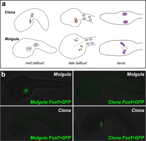
Fig. 9.

Developmental system drift in the cardiopharyngeal mesoderm between Ciona and Molgula. a Diagram comparing differences in morphogenesis of cardiopharyngeal progenitors between Ciona robusta, formerly Ciona intestinalis Type A (top) and Molgula occidentalis (bottom). Mid tailbud stage: lateral view, only one side illustrated. TVCs (green nuclei) separate from their sister anterior tail muscle cells (gray nuclei) and migrate anteriorly and ventrally on each side of the embryo. In M. occidentalis, this migration is more lateral than in C. robusta. Late tailbud stage: ventral view. TVCs divide to give rise to secondary TVCs (yellow) and first heart precursors (red). In C. robusta, these cells form a single cluster at the ventral midline (dotted line), while in M. occidentalis the cells on either side do not meet at the midline. Larva: dorsal view. In C. robusta, atrial siphon muscle precursors (ASMPs, purple) from either side surround an atrial siphon placode (burgundy circle) in the dorsal head epidermis. In M. occidentalis, the future atrial siphon of the juvenile arises from a single primordium that has not yet formed in the larva (dotted burgundy outline). At this stage, the ASMPs of M. occidentalis form two dorsal clusters of cells on either side of the dorsal midline. b Cross-species reporter plasmid assays reveal mutual unintelligibility of orthologous cis-regulatory elements between C. robusta and M. occidentalis. Foxf > GFP reporter plasmids drive identical expression patterns in homologous TVCs when electroporated into embryos of the corresponding species of origin, but are completely non-functional when electroporated into the other species. a Adapted from Kaplan et al. [165]. b Adapted from Stolfi et al. [310]
