Figure 4.

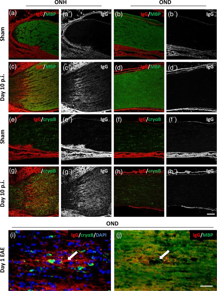
αB‐crystallin expression correlates with areas of anti‐MOG antibody deposition. Immunohistochemistry of optic nerve sections from animals 10 days following immunization with either (a, b, e, f) sham or (c, d, g, h) MOG. Optic nerve head (ONH; a, c, e, g) and distal optic nerve (OND; b, d, f, h) were stained with antibodies against (a–d) IgG deposition and myelin basic protein (MBP); and (e–h) IgG deposition and αB‐crystallin (cryαB). Single channel images of IgG deposition are shown in a'–h'. (i, j) Serial optic nerve sections from a distal segment of optic nerve taken from an animal at day 1 of EAE, stained against (i) IgG deposition, cryαB, and DAPI; and (j) IgG deposition and MBP. White arrow in i and j indicates location of an inflamed blood vessel. Scale bars (h') = 100 μm, (j) = 50 μm
