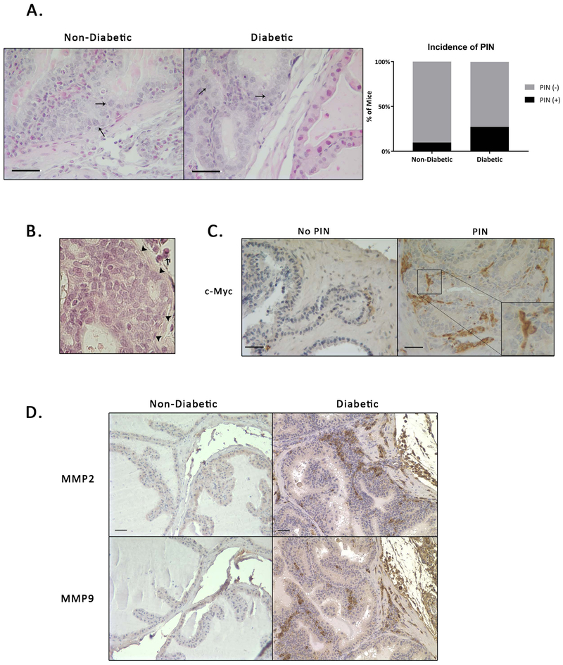
Figure 5. A Subset of Male NOD Mice Develop Areas of PIN-Like Changes.
A) Areas of PIN-like changes in non-diabetic (ND) and diabetic (Di) mice. Arrows indicate enlarged nuclei or increased nuclear-cytoplasmic ratio. B) Representative H&E staining showing no breakdown of extracellular matrix in areas of PIN (arrowheads indicate intact basement membrane, arrow indicates inflammation) C) c-Myc staining revealed positive expression in regions with PIN-Like changes. D) MMP2 and MMP9 staining show increased expression in diabetic mice.

