Abstract
Background:
Germ cell tumors (GCT) are chemosensitive and epidural spinal cord compression (ESCC) from GCT may be amenable to treatment with chemotherapy (CT) only. This retrospective study compares the clinical outcome of GCT patients with ESCC treated with CT or radiotherapy (RT).
Methods:
All patients with a histologic diagnosis of GCT from 1984–2009 were included in this study. Patients with ESCC were identified. Age, clinical features, histology, treatment, and outcome were analyzed.
Results:
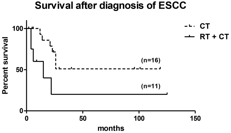
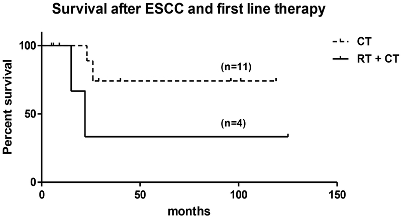
We identified 1734 patients with GCT of whom 29 (1.7%) had ESCC. The median age of these 29 patients was 32 years. The ESCC was treated with CT only in 16 patients, RT+CT in 11, and 2 patients received palliative care only. The ESCC was more extensive in the RT than the CT group. Patients who received RT+CT had a higher proportion of failed prior CT regimens, a higher percentage of non-seminomatous GCT, T-spine involvement, multilevel epidural disease, and bony vertebral metastases. Median overall survival after diagnosis of ESCC was not reached for those treated with CT alone versus 15 month for those receiving RT+CT (p=0.02). There was also a significant difference in survival in patients receiving first line therapy (n=15), where median overall survival was not reached in the CT group (n=11) compared to 22 month in the RT group (n=4) (p=0.04).
Conclusion:
GCT’s rarely involve the epidural compartment. Patients with ESCC who are likely to have chemosensitive disease can receive CT alone as definitive treatment.
Keywords: germ cell tumor, seminoma, non-seminomatous germ cell tumors, epidural cord compression, radiation therapy, chemotherapy
Condensed abstract:
Chemotherapy alone is effective in treating epidural spinal cord compression caused by germ cell tumors with excellent clinical outcomes.
INTRODUCTION
Germ cell tumors (GCT) are uncommon, accounting for only 1%−2% of all malignancies in men, but are the most frequent malignancy in men between the ages of 15 to 35.1–2 Vertebral bone metastasis, spinal cord compression or even spinal cord involvement by GCT is rare and only described in case reports for seminomas3–9 and non-seminomatous germ cell tumors (NSGCT).10–14 In most other solid tumors, ESCC is treated with high-dose corticosteroids with radiation therapy (RT) or surgical decompression. However, ESCC from GCT may be treated effectively by chemotherapy (CT) only.15 The aim of this retrospective analysis is to describe a cohort of patients with ESCC from GCT and to compare survival and neurologic outcome after treating these patients with CT alone in comparison to RT followed by CT (RT+CT).
MATERIALS AND METHODS
The Memorial Sloan-Kettering Cancer Center (MSKCC) clinical database was used to identify patients with a histologic diagnosis of GCT (search terms: “seminoma, NOS”, “seminoma-metastatic NOS”, “seminoma, anaplastic type”, “seminoma-anaplastic,meta”, “spermatocytic seminoma”, “germ cell tumor, nonseminomatous”, “mixed germ cell tumor”, “teratoma”, “choriocarcinoma”, “choriocarcinoma-met”) from 1984–2009. Patients treated before 1984 with chemotherapy alone were already described in an earlier publication and not included into this study.15 Patients with ESCC were identified by ICD-9 code. Age, race/ethnicity, clinical features, histology, treatment, and outcome were analyzed.
The White spinal metastatic cancer grade16 (I: ambulatory; II: non-ambulatory but some motor function; III: paraplegia with no motor function) and the Constans spinal metastases classification17 (I: pain or minor neurological symptoms with normal social and professional activities; II: mild neurological symptoms with normal life but interruption of professional activities; III: moderate neurological symptoms like paraparesis, sphincter disturbances, columnar pain with still active life possible; IV: serious neurological symptoms like paraplegia and complete sphincter deficit; V: medullary syndrome of spinal cord transection) were used to quantify ESCC-related clinical deficits retrospectively. These grades were assigned based on the treating neurologist’s examination done at ESCC diagnosis and during the patient’s treatment. The diagnostic spinal MRI scan was reviewed for all patients in whom films were available, and the degree of ESCC was rated on a scale from 0–3, based on axial T2-weighted MR images; this scale assesses the degree of subarachnoid space obliteration and ESCC and has been used previously.18
Fisher’s exact test was used to calculate significant levels for tumor type, ESCC location and severity on MRI, vertebral versus bone metastases, multilevel disease, prior failed CT, clinical grading scale, and improvement of ESCC on MRI after treatment. Survival was estimated using the Kaplan-Meier method and was compared between patients receiving CT alone versus RT+CT using the log rank test. This study was approved by the MSKCC Institutional Review Board.
RESULTS
We identified 1,734 patients with GCT who met study criteria. In the course of their disease, 393 (23%) patients developed lung metastases, 109 (6%) mediastinal metastases, 147 (8%) GI metastases, 83 (5%) bone metastases, and 29 (1.7%) ESCC of whom 13 had ESCC at presentation. The median age of these 29 patients was 32 years (range 14–59) (Table 1). Ten patients had a seminoma and 19 a NSGCT. The ESCC was treated with CT in 16 patients, 11 received RT+CT, and 2 patients received palliation only (Table 1). Of the 27 patients who received active treatment for ESCC films were available for review in 23 patients, imaging reports confirmed ESCC in the remaining four. Four had ESCC located in the cervical spine, 14 in the thoracic spine, and 9 in the lumbar spine. In 5 of the 27 patients, additional multilevel epidural disease without ESCC was also present (Table 2). 76% of patients had severe ESCC (grade 2–3), 41% had bone metastases, and 44% had a paravertebral mass invading the neuroforamen causing ESCC (Table 3).
Table 1:
Patient Characteristics
| all patients (%) | CT | RT+CT | palliative | |
|---|---|---|---|---|
| # of patients | 29 | 16 | 11 | 2 |
| Age (median, range) | 32 (14–59) | 33 (18–45) | 32 (14–59) | 31 (28–34) |
| Histology | ||||
| Seminoma | 10 (34) | 7 (44) | 3 (27) | 0 |
| NSGCT | 19 (66) | 9 (56) | 8 (73) | 2 (100) |
Table 2:
Clinical Features
| all patients (%) | CT | RT+CT | |
|---|---|---|---|
| # of patients | 27 | 16 | 11 |
| Median time to ESCC (mo) | 15 | 12 | 24 |
| Clinical signs | |||
| Back pain | 26 (96) | 15 (94) | 11 (100) |
| Ambulatory | 23 (85) | 14 (88) | 9 (82) |
| Motor deficits | 10 (37) | 6 (38) | 4 (36) |
| Sensory deficits | 9 (33) | 4 (25) | 5 (45) |
| Reflex abnormalities | 9 (33) | 4 (25) | 5 (45) |
| Incontinence | 5 (19) | 3 (19) | 2 (18) |
| Location | |||
| C-spine | 4 (15) | 3 (19) | 1 (8) |
| T-spine | 14 (52) | 6 (38) | 8 (74) |
| L-spine | 9 (33) | 7 (44) | 2 (18) |
| Multilevel epidural disease | 5 (19) | 0 | 5 (45) |
| Failed prior chemo’s | |||
| 0 | 17 (62) | 12 (75) | 5 (46) |
| 1–2 | 5 (19) | 3 (19) | 2 (18) |
| ≥3 | 5 (19) | 1 (6) | 4 (36) |
Table 3:
Degree, Etiology and clinical Classification of ESCC
| all patients (%) | CT | RT+CT | ||
|---|---|---|---|---|
| # of patients | 27 | 16 | 11 | |
| Degree of ESCC on MRI | ||||
| 0 | 1 (4) | 1 (6) | 0 | |
| 1 | 1 (4) | 1 (6) | 0 | |
| 2 | 11 (40) | 7 (44) | 4 (36) | |
| 3 | 10 (36) | 5 (31) | 5 (45) | |
| No images | 4 (15) | 2 (13) | 2 (18) | |
| ESCC etiology | ||||
| Bone mets | 11 (41) | 5 (31) | 6 (55) | |
| Paravertebral | 12 (44) | 9 (56) | 3 (27) | |
| No images | 4 (15) | 2 (13) | 2 (18) | |
| ESCC on MRI after treatment | ||||
| Improved | 15 (56) | 11 (69) | 4 (36) | |
| Same | 3 (11) | 2 (12) | 1 (9) | |
| Not assessable | 9 (33) | 3 (19) | 6 (55) | |
| WSMCG*: | 21 (78) | 13 (81) | 8 (73) | |
| 4 (15) | 2 (13) | 2 (18) | ||
| 2 (7) | 1 (6) | 1 (9) | ||
| CSMC#: | 9 (33) | 6 (38) | 3 (27) | |
| 9 (33) | 4 (25) | 5 (45) | ||
| 6 (22) | 4 (25) | 2 (18) | ||
| 3 (12) | 2 (13) | 1 (9) | ||
White spinal metastatic cancer grade
Constans spinal metastases classification
Of the patients receiving active treatment 17 (8 with NSGCT) had no prior chemotherapy, 5 (4 with NSGCT) had 1–2 prior chemotherapy regimens, and 5 (all NSGCT) had more than 3 prior regimens. Of the 17 patients with no prior chemotherapy, 13 had ESCC at presentation, 2 had prior RT to the hemipelvis, 1 had orchiectomy only, and 1 denied prior treatment. Patients without ESCC at presentation developed ESCC a median of 86.5 months after initial diagnosis in the CT group compared to a median of 36 months in the RT+CT group (p=0.8).
Patients who received RT+CT had a higher proportion of failed CT regimens (36% versus 6%; p=0.13) which is likely related to the higher percentage of NSGCT (73% versus 56%; p=0.4); T-spine involvement (74% versus 38%; p=0.07); multilevel epidural disease (45% versus 0%; p=0.006); and bony vertebral metastases (55% versus 31%; p=0.21), compared to patients who received CT alone.
The patients receiving CT alone were similar in their clinical presentation to those who received RT+CT with respect to back pain, ambulation, motor deficits, and incontinence, but they had fewer sensory deficits and reflex abnormalities (Table 2). The White spinal metastatic cancer grade had a comparable distribution in both groups, whereas the Constans spinal metastases classification had more patients with severe grades (III, IV) in the CT group (38% versus 27%; p=0.69) (Table 3). The degree of ESCC was similar in the CT and RT+CT groups (grade 3: 31% versus 45%; p=0.69). In the CT group 56% of the ESCC were caused by a paravertebral mass and neuroforaminal infiltration, whereas in the RT+CT group 55% were caused by bony vertebral metastasis (Table 3).
Back pain and ambulation improved in all patients after treatment (Table 4). Incontinence improved in all three patients receiving CT alone whereas neither of the RT+CT patients with incontinence recovered. ESCC on MR imaging improved in 69% of patients receiving CT compared to 36% in the RT+CT group (p=0.13), but pretreatment or follow-up imaging was not available for review in 55% of patients receiving RT+CT compared to 19% in the CT group (Table 3).
Table 4:
Clinical Outcome and Survival
| all patients | CT | RT+CT | |
|---|---|---|---|
| # of patients | 27 | 16 | 11 |
| Symptoms | |||
| Back pain resolved | 26/26 | 16/16 | 11/11 |
| Ambulation restored | 4/4 | 2/2 | 2/2 |
| Incontinence restored | 3/5 | 3/3 | 0/2 |
| Survival (month) | |||
| Median after ESCC | 26 | NR | 15 |
| Median overall | 138 | 138 | 43 |
In the CT alone group, 14 patients (88%) received a platinum-based chemotherapy regimen for their ESCC with bleomycin, etoposide and cisplatin (BEP) being the most commonly used regimen in 9 (56%) patients (Table 5). In the RT+CT group, 8 patients received platinum-based therapy, only 2 of whom received BEP, and one patient each received paclitaxel+iphosphamide, temozolomide, and sunitinib (9% each) after RT (Table 5). Eight patients in the RT+CT group had received chemotherapy for their GCT prior to ESCC development, all of whom received platinum-based chemotherapy (5/8 received BEP (63%)). Fifteen patients received first line chemotherapy for their ESCC of whom 11 received CT alone and 4 RT+CT; all received platinum-based chemotherapy (CT group 9/11 (81%) and RT+CT group 2/4 (50%) received BEP).
Table 5:
Chemotherapeutic Regimen used to Treat ESCC in Patients Receiving Chemotherapy
| CT (%) | RT+CT (%) | |
|---|---|---|
| # of patients | 16 | 11 |
| BEP | 9 (56) | 2 (18) |
| TIP | 3 (19) | 1 (9) |
| VIP | 2 (13) | 1 (9) |
| Paclitaxel+Cisplatin+Ifosphamide | 1 (6) | - |
| CAV | 1 (6) | - |
| Etoposide+Cisplatin | - | 1 (9) |
| Cisplatin+Ifosphamide | - | 1 (9) |
| Paclitaxel+Ifosphamide | - | 1 (9) |
| FOLFOX | - | 2 (18) |
| Temozolomide | - | 1 (9) |
| Sunitinib | - | 1 (9) |
BEP - bleomycin, etoposide and cisplatin
TIP - paclitaxel, ifosfamide, cisplatin
VIP - high dose cisplatin, etoposide, ifosfamide
CAV - cyclophosphamide, doxorubicin, vincristine
FOLFOX - folinic acid+5-FU+oxaliplatin
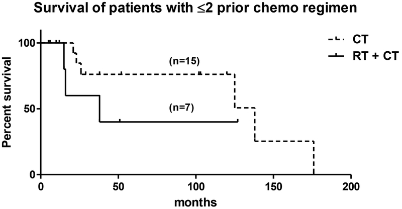
Median survival from GCT was 138 months for those treated with CT alone versus 43 months for those receiving RT+CT (p=0.65). Median survival after diagnosis of ESCC was not reached in the CT group but was only 15 months in the RT group (p=0.02) (Fig. 1). When analyzing the 13 patients who presented with ESCC, all 10 patients who received CT alone responded. Fifteen patients received first line chemotherapy at the time of ESCC; median overall survival for those patients was not reached in the CT group (n=11) compared to 22 months in the RT+CT group (n=4) (p=0.04) (Figure 2). Twenty-two patients received ≤2 prior chemotherapeutic regimens, 15 in the CT and 7 in the RT+CT group. Analysis of these patients, excluding patients heavily pretreated with CT, revealed a median overall survival time of 138 months in the CT and 38 months in the RT+CT group (p=0.23) (Fig. 3).
Figure 1:
Kaplan-Meier survival curve after diagnosis of ESCC. Dashed line represents patients receiving chemotherapy (CT) (n=16), solid line represents patients receiving radiation therapy followed by chemotherapy (RT+CT) (n=11) (p=0.02).
Figure 2:
Kaplan-Meier survival curve after diagnosis of ESCC and treatment with first line therapy. Dashed line represents patients receiving chemotherapy (CT) (n=11), solid line represents patients receiving first line radiation followed by chemotherapy (RT+CT) (n=4) (p=0.04).
Figure 3:
Kaplan-Meier survival curve in patients with ≤2 prior chemotherapy regimens. Dashed line represents patients receiving chemotherapy (CT) (n=15), solid line represents patients receiving radiation therapy followed by chemotherapy (RT+CT) (n=7) (p=0.23).
DISCUSSION
Early detection and aggressive treatment of ESCC are essential for a good neurologic outcome. More than 20,000 new patients per year develop ESCC in the United States.19 Treatment objectives are preservation and improvement of neurologic function, pain relief, and maintenance of spinal stability. The treatment of ESCC is often palliative and includes high dose glucocorticoids, surgical decompression, and RT. A recent randomized trial of ESCC from solid tumors demonstrated a superior outcome with surgical resection followed by RT versus RT alone; however, there were no patients with GCT included in that study.20
ESCC is rare in patients with GCT; in one study of 140 GCT patients, none had ESCC.21 Furthermore, there are no specific data addressing therapy of ESCC in this population. In chemosensitive malignancies, such as GCT, lymphoma, neuroblastoma, or Ewing’s sarcoma, CT could be considered the primary treatment modality for ESCC, providing the expected response would be sufficiently robust as to relieve compression on the spinal cord rapidly.22 Chemotherapy also treats all sites of active disease, sparing patients a delay in systemic treatment and the myelotoxicity of spinal RT. Our patient cohort represents the largest group with ESCC caused by GCT described so far in the literature, and supports the use of chemotherapy as primary treatment for ESCC in these patients.
The first report that cisplatin-based CT could treat ESCC from GCT described three patients from MSKCC.15 Our current series did not include these patients, as they were treated prior to the MRI era. We sought to expand this experience during the period of enhanced diagnostics with spine MRI. In our 27 patients we could demonstrate the effectiveness of CT in the management of GCT with epidural involvement. Patients receiving CT alone recovered their neurologic function, even when there was a high degree of ESCC associated with significant neurologic deficits. These data indicate that CT worked sufficiently quickly that spinal cord compression was relieved promptly and no neurologic deterioration occurred during treatment. In our patient population, RT+CT was used in patients with multilevel epidural disease who were also more heavily pretreated. These patients had more chemoresistent disease and developed ESCC later in the course of their malignancy, so it is unsurprising that patients receiving CT alone had a survival advantage after the diagnosis of ESCC. However, patients who received CT alone as first therapy as well as those who had received ≤2 chemotherapeutic regimens prior to the development of ESCC had longer survival than comparable patients treated with RT+CT; this reached statistical significance only in the first line therapy patients, likely due to the small patient numbers. These patients predictably responded to CT alone for their ESCC and definitive systemic therapy was not delayed for spinal RT which could have compromised overall outcome in patients receiving combined therapy.
There are several limitations to our study, including the small patient number, its retrospective nature and inherent patient selection bias. However, the critical message is clear: patients receiving first line chemotherapy or first or second platinum-based salvage regimens have excellent relief of neurologic symptoms and superior survival when treated with CT alone. This recommendation is based on our limited retrospective data, but a prospective study is unlikely given the rarity of ESCC in GCT. Based on these results, CT alone should be the treatment of choice in newly diagnosed GCT patients who present with ESCC and GCT patients who have received no more than two prior regimens if the disease was previously chemosensitive. Clinical judgement is needed in patients who have received more than two prior chemotherapy regimens, particularly if they have progressed after high dose chemotherapy. In these cases, ESCC is most likely to represent chemoresistant disease, and RT+CT may be the more the more appropriate therapy.
Acknowledgments
This study was not funded by any institution or company.
Footnotes
The authors have nothing to disclose.
Presented in part at the American Society of Clinical Oncology (ASCO) meeting 2010.
REFERENCES
- 1.Horwich A, Shipley J, Huddart R. Testicular germ-cell cancer. Lancet 2006;367(9512):754–65. [DOI] [PubMed] [Google Scholar]
- 2.Devesa SS, Blot WJ, Stone BJ, Miller BA, Tarone RE, Fraumeni JF Jr. Recent cancer trends in the United States. J Natl Cancer Inst 1995;87(3):175–82. [DOI] [PubMed] [Google Scholar]
- 3.Nelson JW, Ruffolo EH. Testicular seminoma as a cause of paraplegia: a case report. J Urol 1966;95(1):70–3. [DOI] [PubMed] [Google Scholar]
- 4.Yee D, Gabos Z, North S, Moore RB. Malignant spinal cord compression secondary to testicular seminoma at the time of initial presentation and at relapse while on surveillance. Can Urol Assoc J 2007;1(1):59–63. [DOI] [PMC free article] [PubMed] [Google Scholar]
- 5.Horvath L, McDowell D, Stevens G, Parkinson R, McCarthy S, Boyer M. Unusual presentations of germ cell tumors. Case 2. Seminoma of the conus medullaris. J Clin Oncol 2001;19(3):911–5. [DOI] [PubMed] [Google Scholar]
- 6.Mackey JR, Venner P. Seminoma with isolated central nervous system relapse, and salvage with craniospinal irradiation. Urology 1998;51(6):1043–5. [DOI] [PubMed] [Google Scholar]
- 7.Kariya S, Kawahara M, Tanimoto I, Matsuki H, Suzumura A. [A case of metastatic extradural seminoma suspected intradural invasion by the measurements of HCG beta concentration in CSF]. Rinsho Shinkeigaku 2000;40(7):722–5. [PubMed] [Google Scholar]
- 8.Gose K, Imajo Y, Takimoto S, Ichiyanagi A, Oshitani T, Kimura S, et al. [Two autopsy cases of intramedullary spinal cord metastasis]. Gan No Rinsho 1984;30(3):319–23. [PubMed] [Google Scholar]
- 9.Umehara F, Okadome T. [A case of the primary mediastinal seminoma invading into the extradural space of the thoracic spine]. Rinsho Shinkeigaku 1990;30(3):304–7. [PubMed] [Google Scholar]
- 10.Lee JK, Kim SH, Kim JH, Kim IY, Kim TS, Jung S, et al. Metastatic spinal cord compression of testicular yolk sac tumor. Childs Nerv Syst 2002;18(3–4):171–4. [DOI] [PubMed] [Google Scholar]
- 11.Dobashi M, Son S, Ikeda M, Fujita T, Matsumoto K, Satoh T, et al. [Three case reports of metastatic germ cell tumors in the lumbar vertebra during first-line chemotherapy]. Hinyokika Kiyo 2008;54(12):803–7. [PubMed] [Google Scholar]
- 12.Kanto S, Tokuyama S, Numahata K, Nakagawa H, Saito S, Arai Y. [Occult lumbar vertebral body metastasis of non-seminomatous germ cell tumor eradicated by radiation and salvage surgery 9 years after initial onset]. Nippon Hinyokika Gakkai Zasshi 2007;98(4):634–7. [DOI] [PubMed] [Google Scholar]
- 13.Colak A, Benli K, Berker M, Onol B. Epidural metastasis of testicular yolk sac tumor: an unusual cause of spinal cord compression. Case report. Pediatr Neurosurg 1991;17(3):139–41. [DOI] [PubMed] [Google Scholar]
- 14.Berglund RK, Lyden SP, Tsai EC, Lieberman I, Klein EA. Nonseminomatous germ cell tumor after chemotherapy with residual mass invading the spine. Eur Urol 2006;50(2):372–4. [DOI] [PubMed] [Google Scholar]
- 15.Cooper K, Bajorin D, Shapiro W, Krol G, Sze G, Bosl GJ. Decompression of epidural metastases from germ cell tumors with chemotherapy. J Neurooncol 1990;8(3):275–80. [DOI] [PubMed] [Google Scholar]
- 16.White WA, Patterson RH Jr. Bergland RM. Role of surgery in the treatment of spinal cord compression by metastatic neoplasm. Cancer 1971;27(3):558–61. [DOI] [PubMed] [Google Scholar]
- 17.Constans JP, de Divitiis E, Donzelli R, Spaziante R, Meder JF, Haye C. Spinal metastases with neurological manifestations. Review of 600 cases. J Neurosurg 1983;59(1):111–8. [DOI] [PubMed] [Google Scholar]
- 18.Wang JC, Boland P, Mitra N, Yamada Y, Lis E, Stubblefield M, et al. Single-stage posterolateral transpedicular approach for resection of epidural metastatic spine tumors involving the vertebral body with circumferential reconstruction: results in 140 patients. Invited submission from the Joint Section Meeting on Disorders of the Spine and Peripheral Nerves, March 2004. J Neurosurg Spine 2004;1(3):287–98. [DOI] [PubMed] [Google Scholar]
- 19.Schiff D Spinal cord compression. Neurol Clin 2003;21(1):67–86, viii. [DOI] [PubMed] [Google Scholar]
- 20.Patchell RA, Tibbs PA, Regine WF, Payne R, Saris S, Kryscio RJ, et al. Direct decompressive surgical resection in the treatment of spinal cord compression caused by metastatic cancer: a randomised trial. Lancet 2005;366(9486):643–8. [DOI] [PubMed] [Google Scholar]
- 21.Mencel PJ, Motzer RJ, Mazumdar M, Vlamis V, Bajorin DF, Bosl GJ. Advanced seminoma: treatment results, survival, and prognostic factors in 142 patients. J Clin Oncol 1994;12(1):120–6. [DOI] [PubMed] [Google Scholar]
- 22.Wong ET, Portlock CS, O’Brien JP, DeAngelis LM. Chemosensitive epidural spinal cord disease in non-Hodgkins lymphoma. Neurology 1996;46(6):1543–7. [DOI] [PubMed] [Google Scholar]


