Abstract
The purpose of the study was to analyze the prevalence of different forms of partial edentulism and the description of the various parameters associated with this disease. Materials and Methods. The study was conducted on a total of 204 subjects who presented themselves at the Clinic of Dental Prosthetics and Oral Rehabilitation Clinic of the Faculty of Dentistry Craiova between October 2015 and June 2016. Results. Of the 204 subjects diagnosed with partial edentulism, 51.47% belonged to the age group of 20-29 years, 52.46% were female and 82.84% came from the urban environment. The most frequent cause of the edentulism was dental caries and their complications. The present study has shown that the partial edentulism forms encountered, especially those of the Kennedy III class, had an increased frequency among the young population, especially in the maxillary arch.
Keywords: Partial edentulism, partial edentulism prevalence, descriptive statistical analysis
Introduction
The term ‘‘edentulism’’ reflects an organ deficiency generally seen in elderly people and has a deep impact on the quality of life [1].
The absence of natural teeth causes functional, esthetic and phonetic problems, and also affects the physiological, biological, social and psychological state of the individuals [2].
Edentulism is a chronic debilitating condition that affects millions of peoples’ ability to function from a physical and psychological standpoint. The edentulous population exists and will continue to increase [3].According to the statistics of Douglass et al. 33.6 million people needed 1 or 2 dentures in the United States in 1991, and, despite dental prevention and the advances in dental technology, this was predicted to increase to 37.9 million in 2020.
The increased longevity of the aged population supports this prediction [4].
Previous studies from different parts of the world have shown that socio-behavioral risk indicators play important roles on edentulism [5]. Potential risk factors for edentulism are low level of education [6], older age [7], gender [8], time since last dental visit [9], health insurance [10], caries severity and periodontal health status [11]. The loss of some or all teeth from the permanent dentition has multiple causes, including both systemic biological and cross-cultural behavioral etiology [12]. Oral health is an integral component of general health and well-being [13].
The retention or loss of permanent teeth is of central importance to an individual’s oral health status and to quality of life. Tooth loss as a measure of oral health has several advantages over other oral health and disease indices, including not only its direct associations with oral function and with overall health and well-being but also the ease by which the presence or absence of teeth can be measured [14]. Furthermore, research has shown that poor oral health in adults may be a risk factor associated with stroke [15], coronary heart disease [16] and acute myocardial infarction [17]. Several studies have concluded that both masticatory efficiency and chewing ability are strongly associated with number and distribution of remaining teeth [18]. Significant associations have been demonstrated between chewing ability and oral-health-related quality of life as well as between chewing ability and cognitive impairment and higher brain functions.Furthermore, impaired dental status has been associated with increased mortality [19]. There is thus still some uncertainty as regards the relationship between chewing ability and dental status, and there is a scarcity of studies in older adults.
The aim of the present study was to analyze the incidence of different forms of partial edentulism and the description of the various parameters associated with it.
Materials and methods
The study was conducted on a total of 204 subjects who presented at the Clinic of Dental Prosthetics and the Oral Rehabilitation Clinic of Craiova Dental Faculty during October 2015-June 2016. The inclusion criteria of the subjects in the study were: adults subjects, aged over 18 years, with one or more partial edentulism forms. The exclusion criteria of the subjects in the study were: subjects aged less than 18 years, with complete dental arches. Each participant in the study was informed on the study objectives and gave his written consent. The present study was approved by the Research Ethics Committee of the University of Medicine and Pharmacy Craiova, according to 1995 Helsinki rules.
The study participants’ data were collected from the interviews and clinical examinations using the questionnaire, anamnesis and clinical examination. The questionnaire included data on systemic illnesses of the subjects. The data obtained by anamnesis included information about age, gender, residence area, profession, and reasons for presentation to the dentist. Clinical examination of each study participant was done according to the methods and the criteria described by the WHO in 1997 [20], under natural daylight in an outdoor setting, using mirrors and ball-ended WHO/CPI periodontal probes (WHO 973/80-Martin, Solingen, Germany). The Kennedy classification of partial edentulism was used to describe the different forms of partial edentulism observed in the study subjects. The Kennedy classification system divides all partially edentulous arches into four basic classes: class I-bilateral edentulous areas located posterior to the remaining natural teeth; class II-a unilateral edentulous area located posterior to the remaining natural teeth; class III-a unilateral edentulous area with natural teeth remaining both anterior and posterior to it; class IV-a single, but bilateral (crossing the midline), edentulous area located anterior to the remaining natural teeth [21].
All the information was noted in individual medical records. The analyzed parameters were: age, gender, residence area, etiology of partial edentulism, number of the absent teeth on the dental arch, partial edentulism topography, Kennedy class of partial edentulism of study, number of participants with prior prosthetic treatments. The collected data were analyzed by descriptive statistics and the correlations between the analyzed parameters were statistically examined using a Chi-square test, with α=5%, the value p<0.05% being considered statistically significant.
Results
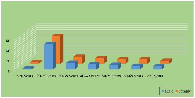
The present study showed that between October 2015 and June 2016, of 204 subjects diagnosed with partial edentulism, 5 participants belonged to the age group of under 20 years representing 2.45% of the total, being the lowest represented category 105 subjects belonged to the age group of20-29 years, representing 51.47% of the total subjects enrolled in the study (Fig.1). Of the 204 subjects diagnosed with partial edentulism, 107 (52.46%) were women and 97 (47.54%) were men, all aged 19-73 years, with an average age of 31 years.The distribution of study participants according to the residence area showed that most of the subjects lived in urban areas (Table 1).
Figure 1.
Distribution of the study subject by age
Table 1.
Distribution of the study subjects according to residence area
|
No. |
Residence area of the study subjects |
Nr. of subjects |
% |
|
1. |
Urban |
169 |
82,84% |
|
2. |
Rural |
35 |
17,16% |
|
3. |
Total number of subjects |
204 |
100% |
From the sample of clinically examined subjects, most of them declared that they suffered teeth loss due to dental extractions as a consequence of complications of carious lesions.
This etiology of partial edentulism was pointed out in 113 of the studied subjects, representing 55.39% of the total.
Another causes of partial edentulism were, also, highlighted (Table 2).
Table 2.
Distribution of subjects according to edentulism etiology
|
No. |
Edentulism etiology |
Nr. of subjects |
% |
|
1. |
Dental caries |
113 |
92% |
|
2. |
Dental inclusion |
1 |
2% |
|
3. |
Anodontia |
24 |
2% |
|
4. |
Periodontitis |
20 |
4% |
|
5. |
Total number of subjects |
204 |
100% |
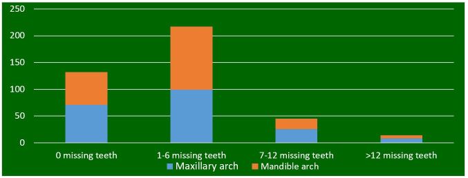
At the level of the maxillary arch, the present study has showed that the number of patients who had 1-6 teeth absent was 99 and 48.52% respectively, and the percentage of patients with 7-12 absent teeth was 12.74%.
In the mandible arch, the statistical study showed that most subjects had 1-6 absent teeth, ie57.84% of the total subjects (Fig.2).
Figure 2.
The distribution of patients in relation to the number of absent teeth on the dental arch
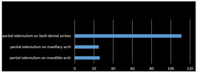
From the analysis of the statistical data, it was observed that most of the examined subjects presented edentulism at the level of both arches, 12.74% had partial edentulism on the mandible arch only, and 25 subjects presented only maxillary edentulism (Fig.3).
Figure 3.
Distribution of subjects according to partial edentulism topography
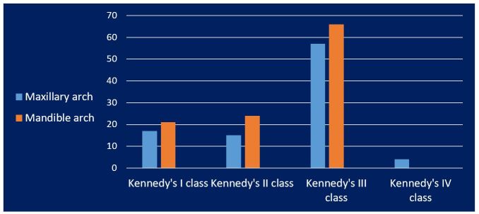
The most frequently Kennedy edentulism class was the third class, both in the mandible and maxillary arches, while the least prevalent form of edentulismwas the forthKennedy class in both arches (Fig.4).
Figure 4.
Distribution of subjects according to the Kennedy class of edentulism
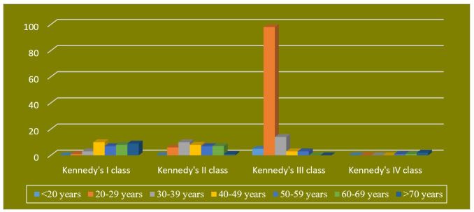
The best-represented edentulism class was Kennedy's 3rd grade, which was especially met in the age group of 20-29 years (Fig.5). A Chi-square test of independence (χ2=6.23, p=0.005) showed a significant correlation between Kennedy’s edentulism classes and age groups (Table 3).
Figure 5.
Distribution of Kennedy edentulism classes in relation to subject age groups
Table 3.
Distribution of study subjects in relation to Kennedy’s classes of edentulism and the age groups
|
No. |
Kennedy Class |
Age group |
Total |
||||||
|
<20 years |
20-29 years |
30-39 years |
40-49 years |
50-59 years |
60-69 years |
>70 years |
|||
|
1 |
I Class |
0 |
1 |
3 |
10 |
7 |
8 |
9 |
38 |
|
2 |
II Class |
0 |
6 |
10 |
8 |
7 |
7 |
1 |
39 |
|
2 |
III Class |
5 |
98 |
14 |
3 |
3 |
0 |
0 |
123 |
|
4 |
IV Class |
0 |
0 |
0 |
0 |
1 |
1 |
2 |
4 |
|
5 |
Total |
5 |
105 |
27 |
21 |
18 |
16 |
12 |
204 |
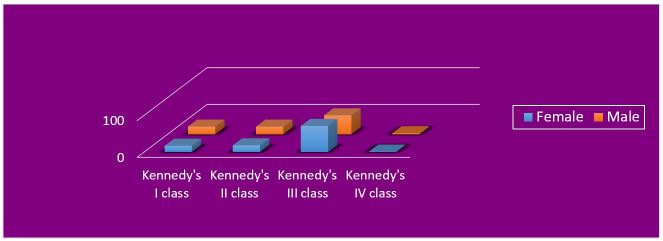
The most frequently class of edentulism was Kennedy’s third class, both in female and male subjects (Fig.6). Statistical analysis using the Chi-square test (χ2=3.75, p=0.250) showed that there is no significant relationship between the Kennedy class of edentulism and the gender of the subjects (Table 4).
Figure 6.
Distribution of Kennedy edentulim classes in relation to subject gender
Table 4.
Distribution of number of subjects in relation to gender and edentulism Kennedy’s classes
|
Kennedy Class/Gender |
Female |
Male |
Total |
|
I Class |
17 |
21 |
38 |
|
II Class |
18 |
21 |
39 |
|
III Class |
71 |
52 |
123 |
|
IV Class |
1 |
3 |
4 |
|
Total |
107 |
97 |
204 |
The statistical study revealed that a number of 30 subjects, ie 14.70% of the total number, presented themselves at the Prosthetic Clinic of the Faculty of Dental Medicine with prosthetic treatments performed.
However, the majority of patients, ie 85.30%, had partial edentulism without any prior prosthetic treatments (Fig7).
Figure 7.
Distribution of subjects in relation to prosthetic treatments
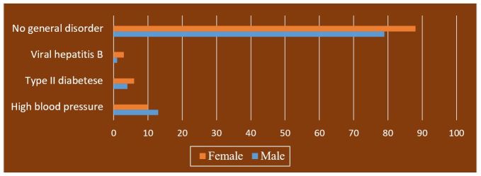
Of the 97 male subjects enrolled in the study, 13 had hypertension, 4 had type II diabetes and only one subject had type B viral hepatitis. Among the female participants, 10 had high blood pressure, 6 had type diabetes II and 3 had type B viral hepatitis (Fig.8). A Chi-square test of independence was calculated. The results (χ2=1.7811, p=0.617) showed that no significant interaction was found between the sex of the subjects and systemic disorders (Table 5).
Figure 8.
Distribution of subjects in relation to the presence of a systemic disorder
Table 5.
Distribution of number of subjects in relation to the presence of a general disorder
|
High blood pressure |
Type II diabetes |
Viral hepatitis B |
No general disorder |
Total |
|
|
Male |
13 |
4 |
1 |
79 |
97 |
|
Female |
10 |
6 |
3 |
88 |
107 |
|
Total |
23 |
10 |
4 |
167 |
204 |
Discussions
Among the various factors studied, age is the key factor found to have significant relationship with occurrence of partial edentulism [2].
As for the age of the study participants who presented a partial edentulism, the majority belonged to the 20-29 age group.
Zaigham AM et al. concluded that with an increase in age, there was an increase in partial edentulism tendency.
In younger age groups, Incidence of partial edentulism was found to be 49% in age group 20-29 years and above 55% in age group 30-39 years [23].
Correlations between subject gender and partial edentulism are another key factor analyzed by different authors.
This study included 204 people aged 18 to 77 years.
Of these, 47.54% were men, and 52.46% were women.
Patel JY, Vohra MY, Hussain JM, et al. argue that there is no significant correlation between genders regarding the appearance of partial edentulism.
However, the authors state that a small number of studies indicate a significant relationship between the gender of the subjects and the different classes of partial edentulism [24]
On the contrary, Abdurahiman VT et al. did not notice a significant gender difference in the appearance of the partial edentulism.
However, the authors observed that men are more prone to the partial edentulism in the posterior maxillary region and women in the posterior mandible region [25].
Zaigam AM et al. found that gender had no correlation with the distribution of partial prostheses, their study comprised 367 patients, of which 157 men and 210 women [23].
Similarly, Abdel Rahman HK et al. showed that gender does not have a statistically significant relationship with the prevalence of different partial edentulism classes [26].
As far as the study participants' background is concerned, the present study shows that most of the patients belonged to the urban area.
According to Daniel M Saman et al. adults with partial edentulism had a probability of 22.6% coming from rural areas and 31.5% being depressed, and adults with total edentulism had a probability of 22.6% to come from the rural area and 31.5% are depressed [27].
Regarding the teeth loss as a cause of partial edentulism, the present study showed that most patients lost their teeth due to dental caries and their complications.
Other authors, such as Vadavadagi SV. et al. showed that most of the subjects surveyed lost their teeth due to periodontal disease (51.04%) and 37.84% of the subjects lost their teeth due to complications of dental caries [28].
Tooth loss and edentulism topography have been the subject of numerous studies.
In the present study it was observed that the prevalence of the partial edentulism was higher in the mandible arch, compared to the maxillary arch, most of the participants having an edentulism form on dental both arches.
These results are in line with the results of Naveed et al., Khalil A. et al. and Patel JY et al., highlighting the fact that partial edentulism frequency was higher in the mandible arch compared to the maxillary arch [29].
On the other hand, Sapkota B et al. show that the partial edentulism was more common in the maxillary arch [30].
In planning the treatment for partial edentulism, the classification on Kennedy's classes plays a vital role in deciding on the type and design of the prosthesis [31].
The present study shows that the most common edentulism class was Kennedy's third class, both in the mandible and maxillary arch, the least frequent being Kennedy's fourth class of edentulism in both arches.
Naveed H et al. observed that the most frequent edentulism class, both in maxillary and mandible arches, was Kennedy's 3rd class. Furthermore, Kennedy's third class with one modification the most common in both arcades [29].
Zaigham AM et al. noticed that the dental arch with a Kennedy class III edentulism was the most common model for the maxillary arch, with the fourth grade being the most rarely encountered [23].
Charyeva OO. et al. found that the most common type of edentulism class was Kennedy class III in both maxillary and mandible arches (41.1%), and the least common was Kennedy's fourth grade for the maxillary (7.1%) and mandible (5.6%) arch [32].
The present study showed that most patients had partial edentulism without prior prosthetic treatments.
According to Hugoson et al. the number of removable partial denture wearers was low but was higher among those aged 80 years (10%) than at age 70 (6%), while the prevalence of subjects with implant treatment was about 13% [33].
Poor oral health has also been associated with lower levels of self-esteem [34], poor mental health [35], and lower quality of life [36].
Data from this study show that most subjects did not have general conditions. However, among those suffering from systemic disorders, the number of men suffering from high blood pressure was significantly higher than the number of women suffering from the same condition, while type II diabetes mellitus and type B viral hepatitis was more commonly encountered in women. Wasif HM. et al., in a study of 570 patients, found that was a significantly higher incidence of cardiovascular disease among males, and a higher percentage of women (56%) who suffered from diabetes. In the same study, they noted that liver disease was more common among men [37].
Conclusions
The present study has shown that the partial edentulism forms, especially those of the Kennedy III class, had an increased frequency among the young population, especially in the maxillary arch.
There were no statistically significant correlations between the type of edentulism and the gender of patients or their residence area.
References
- 1.Özdemir AK, Özdemir HD, Polat NT, Turgut M, Sezer H. The effect of personality type on denture satisfaction. Int. J. Prosthodont. 2006;19(4):364–370. [PubMed] [Google Scholar]
- 2.Scott BJJ, Forgie AH, Davis DM. A study to compare the oral health impact profile and satisfaction before and after having replacement complete dentures constructed by either the copy or the conventional technique. Gerodontology. 2006;23(2):79–86. doi: 10.1111/j.1741-2358.2006.00112.x. [DOI] [PubMed] [Google Scholar]
- 3.White GS. Treatment of the Edentulous Patient. Oral Maxillofacial SurgClin N Am. 2015;27(2):265–272. doi: 10.1016/j.coms.2015.01.005. [DOI] [PubMed] [Google Scholar]
- 4.Douglass CW, Shih A, Ostry L. Will there be a need for complete dentures in the United States in 2020. J Prosthet Dent. 2002;87(1):5–8. doi: 10.1067/mpr.2002.121203. [DOI] [PubMed] [Google Scholar]
- 5.Hugo FN, Hilgert JB, de Sousa MLR, da Silva DD, Pucca Jr GA. Correlates of partial tooth loss and edentulism in the Brazilian elderly. Community Dent. Oral Epidemiol. 2007;35(3):224–232. doi: 10.1111/j.0301-5661.2007.00346.x. [DOI] [PubMed] [Google Scholar]
- 6.Colussi CF, de Freitas. Edentulousness and associated risk factors in a south Brazilian elderly population. Gerodontology. 2007;24(2):93–97. doi: 10.1111/j.1741-2358.2007.00154.x. [DOI] [PubMed] [Google Scholar]
- 7.Fure S, Zickert I. Incidence of tooth loss and dental caries in 60-, 70-and 80-year-old Swedish individuals. Community Dent. Oral Epidemiol. 1997;25(2):137–142. doi: 10.1111/j.1600-0528.1997.tb00911.x. [DOI] [PubMed] [Google Scholar]
- 8.Haikola B, Oikarinen K, Söderholm AL, Remes-Lyly T, Sipilä K. Prevalence of edentulousness and related factors among elderly Finns. J. Oral Rehabil. 2008;35(11):827–835. doi: 10.1111/j.1365-2842.2008.01873.x. [DOI] [PubMed] [Google Scholar]
- 9.Astrøm AN, Haugejorden O, Skaret E, Trovik TA, Klock KS. Oral impacts on daily performance in Norwegian adults: the influence of age, number of missing teeth, and socio-demographic factors. Eur J Oral Sci. 2006;114(2):115–121. doi: 10.1111/j.1600-0722.2006.00336.x. [DOI] [PubMed] [Google Scholar]
- 10.Madlena M, Hermann P, Jahn M, Fejerdy P. Caries prevalence and tooth loss in Hungarian adult population: Results of a national survey. BMC Public Health. 2008;21:8–364. doi: 10.1186/1471-2458-8-364. [DOI] [PMC free article] [PubMed] [Google Scholar]
- 11.Hermann P, Gera I, Borbely J, Fejerdy P, Madlena M. Periodontal health of an adult population in Hungary: findings of a national survey. J. Clin. Periodontol. 2009;36(6):449–57. doi: 10.1111/j.1600-051X.2009.01395.x. [DOI] [PubMed] [Google Scholar]
- 12.Russell SL, Gordon S, Lukacs JR, Kaste LM. Sex/Gender differences in tooth loss and edentulism: historical perspectives, biological factors, and sociologic reasons. Dent Clin North Am. 2013;57(2):317–337. doi: 10.1016/j.cden.2013.02.006. [DOI] [PubMed] [Google Scholar]
- 13.Satcher D, Nottingham JH. Revisiting Oral Health in America: A Report of the Surgeon General. American Journal of Public Health. 2017;107(S1):S32–S33. doi: 10.2105/AJPH.2017.303687. [DOI] [PMC free article] [PubMed] [Google Scholar]
- 14.Russell SL, Gordon S, Lukacs JR, Kaste LM. Sex/Gender differences in tooth loss and edentulism: historical perspectives, biological factors, and sociologic reasons. Dent Clin North Am. 2013;57(2):317–337. doi: 10.1016/j.cden.2013.02.006. [DOI] [PubMed] [Google Scholar]
- 15.Elter JR, Offenbacher S, Toole JF, Beck JD. Relationship of periodontal disease and edentulism to stroke/TIA. J Dent Res. 2003;82(12):998–1001. doi: 10.1177/154405910308201212. [DOI] [PubMed] [Google Scholar]
- 16.Hung HC, Joshipura KJ, Colditz G, Manson JE, Rimm EB, Speizer FE, Willett WC. The association between tooth loss and coronary heart disease in men and women. J Public Health Dent. 2004;64(4):209–215. doi: 10.1111/j.1752-7325.2004.tb02755.x. [DOI] [PubMed] [Google Scholar]
- 17.Willershausen B, Kasaj A, Willershausen I, Zahorka D, Briseño B, Blettner M, Genth-Zotz S, Münzel T. Association between chronic dental infectionand acute myocardial infarction. J Endod. 2009;35(5):626–630. doi: 10.1016/j.joen.2009.01.012. [DOI] [PubMed] [Google Scholar]
- 18.Naka O, Anastassiadou V, Pissiotis A. Association between functional tooth units and chewing ability in older adults: a systematic review. Gerodontology. 2014;31(3):166–177. doi: 10.1111/ger.12016. [DOI] [PubMed] [Google Scholar]
- 19.Schwahn C, Polzer I, Haring R, Dörr M, Wallaschofski H, Kocher T, Mundt T, Holtfreter B, Samietz S, Völzke H, Biffar R. Missing, unreplaced teeth and risk of all cause and cardiovascular mortality. Int J Cardiol. 2013;20; 167(4):1430–1437. doi: 10.1016/j.ijcard.2012.04.061. [DOI] [PubMed] [Google Scholar]
- 20. World Health Organization . Oral Health Surveys: Basic Methods . 4 . Geneva, England : 1997 . [Google Scholar]
- 21.Al-Johany SS, Andres C. ICK Classification System for Partially Edentulous Arches. J Prosthodont. 2008;17(6):502–507. doi: 10.1111/j.1532-849X.2008.00328.x. [DOI] [PubMed] [Google Scholar]
- 22.Sadiq WM, Idowu AT. Removable Partial denture design: A study of a selected population in Saudi Arabia. J Contemp Dent Pract. 2002;3(4):1–11. [PubMed] [Google Scholar]
- 23.Zaigham AM, Muneer MU. Pattern of partial edentulism and its association withage and gender. Pak Oral Dent J. 2010;30(1):260–263. [Google Scholar]
- 24.Patel JY, Vohra MY, Hussain JM. Assessment of Partially edentulous patients based on Kennedy’s classification and its relation with Gender Predilection. International Journal of ScientificStudy. 2014;2(6):32–36. [Google Scholar]
- 25.Abdurahiman VT, Kahdar MA, Jolly SJ. Frequency of partial edentulism and awareness to restore the same: A Cross sectional study in the age group of 18-25 years among Kerala student population. J Indian ProsthodontSoc. 2013;13(4):461–465. doi: 10.1007/s13191-012-0246-2. [DOI] [PMC free article] [PubMed] [Google Scholar]
- 26.Abdel-Rahman HK, Tahir CD, Saleh MM. Incidence of Partial edentulism and its relation with age and gender. Zanco J Med Sci. 2013;17:463–470. [Google Scholar]
- 27.Nalcacı R, Erdemir EO, Baran I. Evaluation of the oral health status of the people aged 65 years and over living in near rural district of Middle Anatolia,Turkey. Arch. Gerontol. Geriatr. 2007;45(1):55–64. doi: 10.1016/j.archger.2006.09.002. [DOI] [PubMed] [Google Scholar]
- 28.Vadavadagi SV, SrinivasaH H, Goutham GB, Hajira N, Lahari M, Prasantha Reddy GT. Partial Edentulism and its Association with Socio-Demographic Variables among Subjects Attending Dental Teaching Institutions, India. J Int Oral Health. 2015;7(Suppl 2):60–63. [PMC free article] [PubMed] [Google Scholar]
- 29.Naveed H, Aziz MS, Hassan A, Khan W, Azad AA. Patterns of Partial Edentulismamong armed forces personnel reporting at armed forces institute of dentistryPakistan. Pak Oral Dent J. 2011;31(1):217–221. [Google Scholar]
- 30.Sapkota B, Adhikari B, Upadhaya C. A Study of Assessment of Partialedentulous patients based on Kennedy’s classification at DhulikhelHospitalKathmandu University Hospital. Kathmandu Univ Med J. 2013;44(4):325–327. doi: 10.3126/kumj.v11i4.12542. [DOI] [PubMed] [Google Scholar]
- 31.König J, Holtfreter B, Kocher T. Periodontal health in Europe: Future trends based on treatment needs and the provision of periodontal services-position paper 1. Eur. J. Dent. Educ. 2010;14Suppl1:4–24. doi: 10.1111/j.1600-0579.2010.00620.x. [DOI] [PubMed] [Google Scholar]
- 32.Charyeva OO, Altynbekov KD, Nysanova BZ. Kennedy classification andtreatment options: A study of partially edentulous patients being treated in a specialized prosthetic clinic. J Prosthodont. 2012;21(3):177–180. doi: 10.1111/j.1532-849X.2011.00809.x. [DOI] [PubMed] [Google Scholar]
- 33.Hugoson A, Koch G, Gothberg C, Helkimo AN, Lundin SA, Norderyd O. Oral health of individuals aged 3-80 years in Jonkoping, Sweden during 30 years (1973-2003). II. Review of clinical and radiographic findings. Swed Dent J. 2005;29:139–155. [PubMed] [Google Scholar]
- 34.Gift HC, Reisine ST, Larach DC. The social impact of dental problems and visits. Am J Public Health. 1992;82:1663–1668. doi: 10.2105/ajph.82.12.1663. [DOI] [PMC free article] [PubMed] [Google Scholar]
- 35.Quine S, Morrell S. Hopelessness, depression and oral health concerns reported by community dwelling older Australians. Community Dent Health. 2009;26:177–182. [PubMed] [Google Scholar]
- 36. Locker D . Concepts of oral health, disease and the quality of life . In: Slade GD , editor. Measuring oral health and quality of life . North Carolina : Chapel Hill ; 1997 . pp. 11 – 23 . [Google Scholar]
- 37.Wasif HM HM, Tanwir F, Nawaz M. Association of Systemic Diseases on Tooth Loss and Oral Health. J Biomedical Sci. 2015;4:1–7. [Google Scholar]


