Abstract
Neuroendocrine tumors (NETs) of gastrointestinal (GI) tract and lungs are a rare variety of tumors but given their indolent nature are quite prevalent. These tumors are mostly malignant in nature and are often diagnosed in advanced stages. GI tracts are the most common sites of NETs followed by lungs, thymus, and other less common sites being ovaries, testis, and hepatobiliary system. Nuclear medicine imaging modalities include 68Ga-DOTANOC positron emission tomography/computed tomography (PET/CT) which is sensitive for low-grade NETs and 18F-fluorodeoxyglucose (FDG) PET/CT which is more valuable for high-grade NETs. However, intermediate-grade NETs are equally sensitive to both 68Ga-DOTANOC PET/CT and 18F-FDG PET/CT.
Keywords: 18F-fluorodeoxyglucose positron emission tomography/computed tomography, 68Ga-DOTANOC positron emission tomography/computed tomography, neuroendocrine tumors
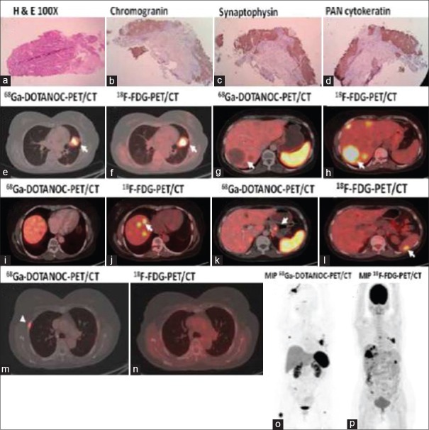
A 55-year-old female consulted the pulmonary medicine department with chief complaints of obstructive pneumonia, chest pain, dyspnea, and cough. Computed tomography (CT)-guided biopsy of the left lung mass showed tissue consistent with Grade 2 neuroendocrine tumor (NET) [Figure 1a] which was immunopositive for chromogranin [Figure 1b], synaptophysin [Figure 1c], and cytokeratin [Figure 1d]. She was subsequently referred to the nuclear medicine department for whole-body 68Ga-DOTANOC positron emission tomography/computed tomography (PET/CT) and 18F-fluorodeoxyglucose (FDG) PET/CT scans to detect the extent of disease. Grade-2 NET lung detected on biopsy (primary) showed positive uptake on both 68Ga-DOTANOC PET/CT [arrow, Figure 1e] and 18F-FDG PET/CT [arrow, Figure 1f] scans. However, multiple metastatic lesions in the liver showed no somatostatin receptor expression as evidenced by negative 68Ga-DOTANOC PET/CT [arrow, Figure 1g and i] but showed avid lesions on 18F-FDG PET/CT [arrow, Figure 1h and j], proving it to be as poorly differentiated metastases. Similarly, another solitary metastatic lesion noted in the pancreas was positive on 68Ga-DOTANOC PET/CT scan [arrow, Figure 1k] but showed no radiotracer uptake on 18F-FDG PET/CT [Figure 1l], implying it to be a well-differentiated metastatic lesion. Another lesion noted in the spleen was positive on 18F-FDG PET/CT [arrow, Figure 1l], but no tracer uptake was observed on 68Ga-DOTANOC PET/CT scan [Figure 1k], consistent with poorly differentiated uptake pattern. Multiple bone metastases were positive on 68Ga-DOTANOC PET/CT [arrow, Figure 1m] but negative on 18F-FDG PET/CT scan [Figure 1n], which implied it to be well differentiated. Figure 1o and p depict the maximum intensity projection images of 68Ga-DOTANOC PET/CT [Figure 1o] and 18F-FDG PET/CT [Figure 1p]. Bronchial carcinoids are rare well-differentiated NET subclassified into typical and atypical carcinoids.[1] Using 68Ga-DOTANOC PET/CT and 18F-FDG PET/CT scan, we can differentiate between various grades of NETs.[2] 68Ga-DOTANOC PET/CT is the initial evaluation of choice for detection of primary sites for patients with metastatic NETs of unknown origin as well as well-differentiated NETs.[3] 18F-FDG PET/CT also has an important role in managing patients with aggressive and high-grade NETs owing to its high prognostic value, and further treatment change is based on this poor differentiation of the tumor. 68Ga-DOTANOC PET-CT alone at the best is helpful in delineating disease extent. The evaluation of glycolytic metabolism by 18F-FDG is potentially useful in identifying high-risk patients with aggressive neuroendocrine disease associated with a poor outcome.[4]68-Ga DOTANOC PET/CT and 18F-FDG PET/CT have a complementary role in patients who have aggressive disease as it helps in demonstrating the total disease burden and stratify them to proper therapeutic groups.[5] Dual-tracer imaging with 68-Ga-DOTANOC and 18-F-FDG PET-CT can have a prognostic value in metastatic NETs as positive somatostatin receptor expression with negative FDG uptake exhibits a longer progression-free survival.[6] In such patients, somatostatin analogs can be used as first-line agent, while high FDG uptake warrants treatment with chemotherapy.[7,8] In this case, splenic lesion was positive on 18-F FDG PET/CT and negative on 68Ga DOTANOC PET/CT. One must be careful in reporting splenic lesion on 68Ga-DOTANOC PET/CT as such lesions could be missed due to high physiological splenic DOTANOC uptake. This case highlights the fact that in a metastatic NET, various grades of differentiation can be present at a given point of time. Hence, imaging with 18-F-FDG PET/CT and 68-Ga-DOTANOC PET/CT can give a complete picture about the disease status and should be used in routine for better management of patients.
Figure 1.
(a-d) Grade II neuroendocrine tumor immunopositive for chromogranin, synaptophysin cytokeratin, (e and f) neuroendocrine tumor Grade II lung, both DOTANOC and fluorodeoxyglucose positive, (g and i) DOTANOC-negative liver lesions, (h and j) fluorodeoxyglucose-positive liver lesions, (k) DOTANOC-positive pancreas lesion and DOTANOC-negative spleen lesion, (l) fluorodeoxyglucose-negative pancreas lesion and fluorodeoxyglucose-positive spleen lesion, (m) DOTANOC-positive bone metastases, (n) fluorodeoxyglucose-negative bone metastases, (o) maximum intensity projection image of 68-Ga DOTANOC positron emission tomography/computed tomography, (p) maximum intensity projection image of 18-F-fluorodeoxyglucose positron emission tomography/computed tomography
Declaration of patient consent
The authors certify that they have obtained all appropriate patient consent forms. In the form the patient(s) has/have given his/her/their consent for his/her/their images and other clinical information to be reported in the journal. The patients understand that their names and initials will not be published and due efforts will be made to conceal their identity, but anonymity cannot be guaranteed.
Financial support and sponsorship
Nil.
Conflicts of interest
There are no conflicts of interest.
References
- 1.Ambrosini V, Castellucci P, Rubello D, Nanni C, Musto A, Allegri V, et al. 68Ga-DOTA-NOC: A new PET tracer for evaluating patients with bronchial carcinoid. Nucl Med Commun. 2009;30:281–6. doi: 10.1097/MNM.0b013e32832999c1. [DOI] [PubMed] [Google Scholar]
- 2.Lococo F, Treglia G. Which is the best strategy for diagnosing bronchial carcinoid tumours? The role of dual tracer PET/CT scan. Hell J Nucl Med. 2014;17:7–9. doi: 10.1967/s002449910111. [DOI] [PubMed] [Google Scholar]
- 3.Pruthi A, Pankaj P, Verma R, Jain A, Belho ES, Mahajan H, et al. Ga-68 DOTANOC PET/CT imaging in detection of primary site in patients with metastatic neuroendocrine tumours of unknown origin and its impact on clinical decision making: Experience from a tertiary care centre in India. J Gastrointest Oncol. 2016;7:449–61. doi: 10.21037/jgo.2016.01.06. [DOI] [PMC free article] [PubMed] [Google Scholar]
- 4.Panagiotidis E, Bomanji J. Role of 18F-fluorodeoxyglucose PET in the study of neuroendocrine tumors. PET Clin. 2014;9:43–55. doi: 10.1016/j.cpet.2013.08.008. [DOI] [PubMed] [Google Scholar]
- 5.Naswa N, Sharma P, Gupta SK, Karunanithi S, Reddy RM, Patnecha M, et al. Dual tracer functional imaging of gastroenteropancreatic neuroendocrine tumors using 68Ga-DOTA-NOC PET-CT and 18F-FDG PET-CT: Competitive or complimentary? Clin Nucl Med. 2014;39:e27–34. doi: 10.1097/RLU.0b013e31827a216b. [DOI] [PubMed] [Google Scholar]
- 6.Bahri H, Laurence L, Edeline J, Leghzali H, Devillers A, Raoul JL, et al. High prognostic value of 18F-FDG PET for metastatic gastroenteropancreatic neuroendocrine tumors: A long-term evaluation. J Nucl Med. 2014;55:1786–90. doi: 10.2967/jnumed.114.144386. [DOI] [PubMed] [Google Scholar]
- 7.Panagiotidis E, Alshammari A, Michopoulou S, Skoura E, Naik K, Maragkoudakis E, et al. Comparison of the impact of 68Ga-DOTATATE and 18F-FDG PET/CT on clinical management in patients with neuroendocrine tumors. J Nucl Med. 2017;58:91–6. doi: 10.2967/jnumed.116.178095. [DOI] [PubMed] [Google Scholar]
- 8.Has Simsek D, Kuyumcu S, Turkmen C, Sanlı Y, Aykan F, Unal S, et al. Can complementary 68Ga-DOTATATE and 18F-FDG PET/CT establish the missing link between histopathology and therapeutic approach in gastroenteropancreatic neuroendocrine tumors? J Nucl Med. 2014;55:1811–7. doi: 10.2967/jnumed.114.142224. [DOI] [PubMed] [Google Scholar]


