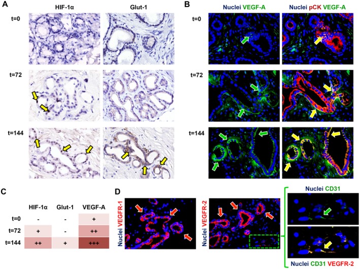
Figure 4.

Activation of angiogenic factors during incubation. (A) Immunohistochemistry for the hypoxic response markers HIF‐1α and Glut‐1 showed up‐regulation during incubation (yellow arrows). (B) Correspondingly, VEGF‐A expression increased over time (green arrows) and was expressed by pCK‐positive PBG cells (yellow arrows). (C) Semiquantitative evaluation of angiogenic factor expression in PBG cells showed up‐regulation of HIF‐1α, Glut‐1, and VEGF‐A over time. (D) VEGF receptors VEGFR‐1 and VEGFR‐2 were expressed by PBG cells (red arrows) and CD31‐positive endothelial cells (green arrow) expressed VEGFR‐2 at their cell membrane. Area in the box is magnified on the right and separate channels are provided. (A‐C) Original magnification ×40. Abbreviations: CD31, cluster of differentiation 31; pCK, pan cytokeratin.
