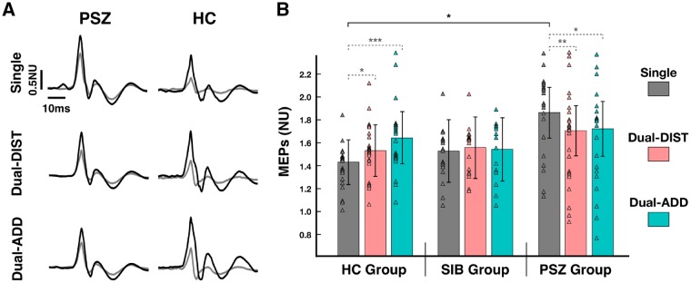
Figure 4.
Cortical excitability. (A) Raw data of MEPs recorded during the visuomotor grip force-tracking task (hold period) for a patient with schizophrenia (PSZ) and a healthy control subject (HC). Example trials show an unconditioned MEP (dark line) and a conditioned MEP (grey line) of the 1DI for (i) the single-task force-tracking condition (Single) and the dual-task condition with (ii) dual-task distraction trial (Dual-DIST) and (iii) dual-task addition trial (Dual-ADD). (B) Mean normalized amplitude of MEPs for the hold period (estimated marginal mean ± vertical bars: 95% CI) during Single (grey), Dual-DIST (pink) and Dual-ADD (cyan) trials for the three groups: patients with schizophrenia, healthy controls and siblings (SIB). Triangles represent data points of the individual subjects in each group and condition. Significant differences (LSD fisher post hoc tests for between-group comparisons are shown as horizontal black brackets and within-group comparisons as horizontal grey dashed bracket with: *P < 0.05; **P < 0.01, ***P < 0.001. For clarity, significant differences for between-group comparisons are only indicated between patients and healthy controls. Post hoc tests for between-group comparisons (not indicated) revealed that patients with schizophrenia showed an increased excitability only in single-task condition compared to healthy controls (patients with schizophrenia versus healthy controls: P = 0.02), this was not significantly different compared to siblings (patients with schizophrenia versus siblings: P = 0.08).

