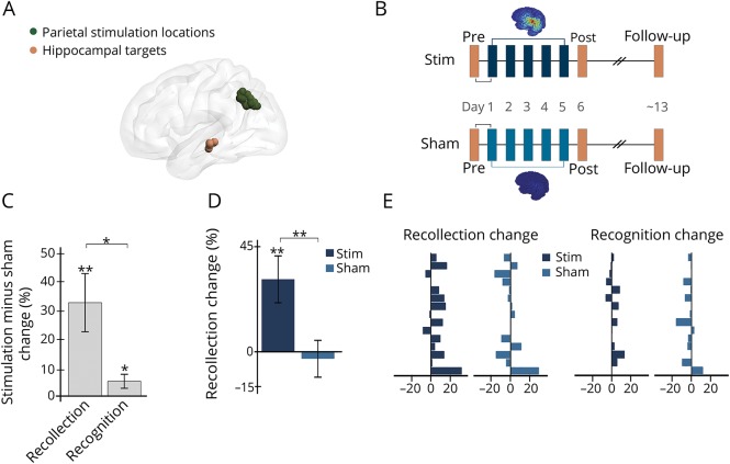
Figure 1. Stimulation increased recollection accuracy.
(A) Participant-specific stimulation locations (one sphere shown per participant) were selected based on high seed-based resting-state fMRI connectivity with hippocampal target locations (one sphere shown per participant). (B) Before and approximately 24 hours after 5 consecutive daily sessions of full-intensity or sham stimulation, participants completed fMRI memory assessments. Stimulation and sham were administered in all participants in counterbalanced order using a within-subjects crossover design. Approximately 1 week following the post-stim and post-sham sessions, participants returned for follow-up assessments. Stimulation-induced electrical field for each stimulation condition is displayed for a representative participant with warmer colors representing peak intensity (range: 1–119 V/m). (C) Effects of stimulation on recollection and on recognition calculated for stimulation vs sham at the 24-hour assessment. (D) Recollection changes due to stimulation and sham. (E) Each bar represents a single-participant change in recollection and recognition for stimulation and sham, demonstrating consistent improvement due to stimulation particularly for recollection. Error bars indicate SEM. *p ≤ 0.05, **p ≤ 0.01.

