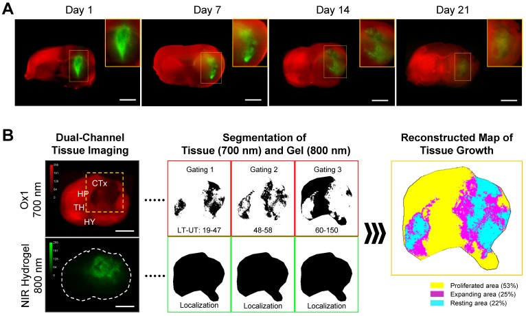
Figure 4.
Dual-channel imaging of brain tissue growth and NIR hydrogel. (A) NIR hydrogel (20 µL) was injected into the brain of animals and Ox1 (100 nmol) was administered to the same animal an hour prior to the dual-channel imaging. Brain tissue ingrowth (red) and NIR hydrogel (green) degradation observed in the merged image. Sample thickness = 2 mm. (B) Spatial segmentation analysis of the NIR image for the quantification and visualized dynamic color mapping of the tissue ingrowth. Scale bars = 2.5 mm.

