Short abstract
Improvement in breast cancer screening technology has increased the detection of architectural distortion, which can often indicate underlying malignancy; however, there are also many benign causes of architectural distortion. We present a case of architectural distortion caused by cyst aspiration, representing a novel, benign cause.
Keywords: Breast, mammography, tomosynthesis, cyst aspiration, architectural distortion
Introduction
Historically, standard two-dimensional mammography has been the mainstay of early breast cancer detection (1). Improvement in breast cancer screening technology includes digital breast tomosynthesis, FDA-approved in 2011, which allows for reconstruction of 1-mm slices through the breast volume (2). Tomosynthesis reduces recall rate, increases the cancer detection rate, and is significantly better at visualizing architectural distortion (AD) over conventional mammography (1,3–6).
AD refers to multiple converging straight lines within the breast parenchyma, without a definable mass lesion (7). Although AD can indicate underlying malignancy, there are also many benign causes that have been reported in the literature (8,9). We present a case of AD caused by cyst aspiration, which represents a novel, benign cause of AD not previously described.
Case report
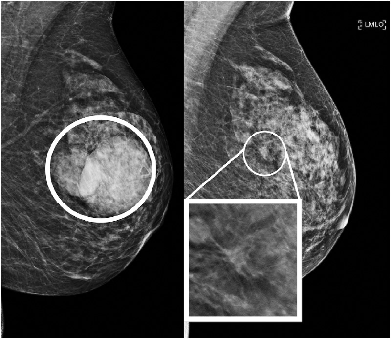
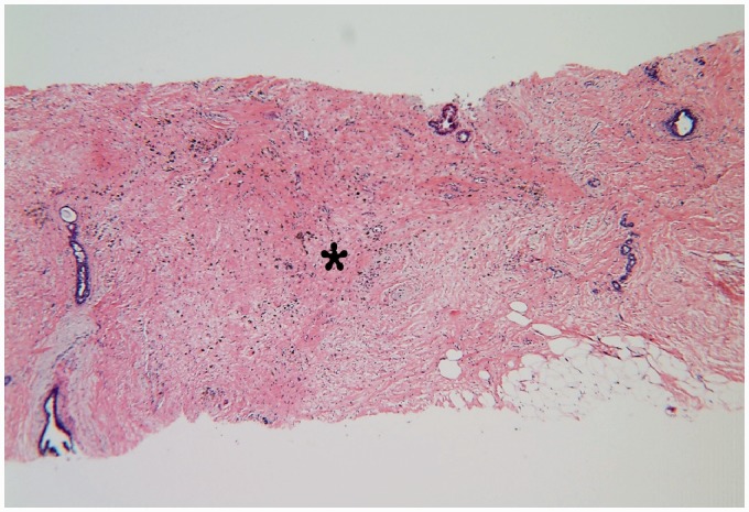
A woman in her 50s presented with bilateral lumps and pain; she was found to have four cysts. She subsequently underwent cyst aspiration of all four cysts in 2016 for symptomatic relief utilizing an 18-G needle with 12-mL syringe and 1% lidocaine for local anesthetic. Two years later, the patient returned for her first routine screening mammogram after her aspiration, resulting in a recall (BI-RADS 0) for bilateral AD at the site of prior cyst aspirations (Figs. 1 and 2). The AD persisted bilaterally on diagnostic mammography and was without sonographic correlates on ultrasound. The diagnostic study was coded BI-RADS 4, biopsy recommended, for suspicious bilateral AD. Targeted bilateral tomosynthesis-guided needle biopsy using a Mammotome Revolve 8-G vacuum needle mated to a Hologic Affirm prone biopsy table was performed, which revealed focal fibrosis and hemosiderin laden macrophages on pathology (Figs. 3 and 4). The pathology results were determined to be concordant with radiologic findings, given the histologic findings and characteristic location overlying areas of prior cyst aspiration.
Fig. 1.
Mediolateral oblique view of the left breast from 2016 (left) and 2018 (right) showing interval development of AD at the site of cyst aspiration.
Fig. 2.
Mediolateral oblique view of the right breast from 2016 (left) and 2018 (right) showing interval development of AD at the site of cyst aspiration.
Fig. 3.
H&E stain of right breast tissue core shows stromal fibrosis (4×).
Fig. 4.
High power shows hemosiderin laden macrophages (arrow) associated with fibrosis (20×).
Discussion
Tomosynthesis has been shown to improve sensitivity in detecting AD, which has contributed to an overall increase in screening performance (5,10). Once detected, further evaluation is needed to exclude malignancy as the positive predictive value of AD for malignancy has been reported at 74.5% (11). Ultrasound is the first-line imaging study for attempt in identifying a target for needle biopsy. Even if ultrasound is negative, tomosynthesis-guided needle biopsy or wire localization would be warranted for tissue diagnosis. Before the development of tomosynthesis-guided procedures, magnetic resonance imaging (MRI) for evaluation of a correlate was often performed (12).
Malignant causes of AD are well documented and are most common with invasive ductal or lobular carcinoma and have also been described as a prominent feature of tubular carcinomas (8).
The most common benign causes of AD include radial scars, sclerosing adenosis, fat necrosis, or scarring related to surgery or needle biopsies (8,9). A review of the literature has shown no previously reported association between cyst aspiration and AD. The importance of this case report is to highlight cyst aspiration as a potential benign concordant cause of AD and show that it can inform decisions regarding post-biopsy radiologic–pathologic correlation. The benign pathology findings of fibrosis should be corroborated to the location of prior aspiration. It is currently unclear which patient-related factors or cyst features portend eventual formation of AD. In our patient, four total cysts were aspirated, but only two sites developed AD. The presence of hemosiderin laden macrophages in the fibrosis may suggest that hemorrhage in the cyst, either from the needle introduction or cyst inflammation, may play a factor in the development of post-cyst aspiration AD.
In conclusion, with cases of AD, benign and malignant etiologies must be considered. Prior cyst aspiration, with correlating location and pathologic findings is a concordant result for benign AD, mimicking other more common causes of distortion such as malignancy, radial scar, sclerosing adenosis, fat necrosis, or post-surgical scarring.
Declaration of conflicting interests
The authors declared no potential conflicts of interest with respect to the research, authorship, and/or publication of this article.
Funding
The authors received no financial support for the research, authorship, and/or publication of this article.
References
- 1.Vijapura C, Yang L, Xiong J, et al. Imaging features of nonmalignant and malignant architectural distortion detected by tomosynthesis. AJR Am J Roentgenol 2018; 211:1397–1404. [DOI] [PubMed] [Google Scholar]
- 2.Dobbins JT. Tomosynthesis imaging: at a translational crossroads. Med Phys 2009; 36:1956–1967. [DOI] [PMC free article] [PubMed] [Google Scholar]
- 3.Phantana-angkool A, Forster MR, Warren YE, et al. Rate of radial scars by core biopsy and upgrading to malignancy or high-risk lesions before and after introduction of digital breast tomosynthesis. Breast Cancer Res Treat 2019; 173:23–29. [DOI] [PubMed] [Google Scholar]
- 4.Bahl M, Lamb LR, Lehman CD. Pathologic outcomes of architectural distortion on digital 2D versus tomosynthesis mammography. AJR Am J Roentgenol 2017; 209:1162–1167. [DOI] [PubMed] [Google Scholar]
- 5.Dibble EH, Lourenco AP, Baird GL, et al. Comparison of digital mammography and digital breast tomosynthesis in the detection of architectural distortion. Eur Radiol 2018; 28:3–10. [DOI] [PubMed] [Google Scholar]
- 6.Partyka L, Lourenco AP, Mainiero MB. Detection of mammographically occult architectural distortion on digital breast tomosynthesis screening: initial clinical experience. AJR Am J Roentgenol 2014; 203:216–222. [DOI] [PubMed] [Google Scholar]
- 7.Sickles EA, D’Orsi CJ, Bassett LW, et al. ACR BI-RADS® Mammography. In: ACR BI-RADS® Atlas, Breast Imaging Reporting and Data System. Reston, VA, American College of Radiology; 2013.
- 8.Boyer B, Russ E. Anatomical-radiological correlations: architectural distortions. Diagn Interv Imaging 2014; 95:134–140. [DOI] [PubMed] [Google Scholar]
- 9.Gaur S, Dialani V, Slanetz PJ, et al. Architectural distortion of the breast. AJR Am J Roentgenol 2013; 201:W662–670. [DOI] [PubMed] [Google Scholar]
- 10.Rose SL, Shisler JL. Tomosynthesis impact on breast cancer screening in patients younger than 50 years old. AJR Am J Roentgenol 2018; 210:1401–1404. [DOI] [PubMed] [Google Scholar]
- 11.Bahl M, Baker JA, Kinsey EN, et al. Architectural distortion on mammography: correlation with pathologic outcomes and predictors of malignancy. AJR Am J Roentgenol 2015; 205:1339–1345. [DOI] [PubMed] [Google Scholar]
- 12.Durand MA, Wang S, Hooley RJ, et al. Tomosynthesis-detected architectural distortion: management algorithm with radiologic-pathologic correlation. Radiographics 2016; 36:311–321. [DOI] [PubMed] [Google Scholar]


