Figure 2.
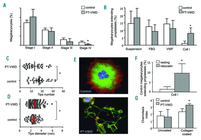
Proplatelet formation from human megakaryocytes. (A) Megakaryocyte percentage and maturation. The percentage of CD41+ cells at day 14 of culture was measured by flow cytometry. Maturation of megakaryocytes was determined by fluorescence microscopy based on ploidy, cell diameter, and CD41 expression. For each sample, at least 100 megakaryocytes were evaluated. Data represent mean±Standard Deviation (SD) for five controls and for the platelet-von Willebrand disease (PT-vWD) patient on five different occasions; *P<0.05 vs. control. (B) Percentage of megakaryocytes-extending proplatelets in suspension, or onto glass coverslips coated with fibrinogen (FBG), vWF or type I collagen (Coll I). Data represent mean±Standard Error of Mean (SEM) of five controls and of five different preparations from the PT-vWD patient (*P<0.05 vs. controls). (C) Representative images of control and PT-vWD megakaryocytes plated on type I collagen. Scale bars=20 μm. β1 tubulin is stained green (Alexa Fluor® 488 Goat Anti-Rabbit IgG; Molecular Probes, Life Technologies, Milan, Italy), polymerized actin is stained red (rhodamine-phalloidine; Molecular Probes), and nuclei are stained blue with Hoechst. Specimens were mounted with the ProLong Antifade medium (Molecular Probes), analyzed at room temperature with a Carl Zeiss Axio Observer.A1 fluorescence microscope (Carl Zeiss Inc., Oberkochen, Germany) using a 63×/1.4 Plan-Apochromat oil-immersion objective and images acquired using the AxioVision software (Carl Zeiss Inc.). All polynucleated cells extending protrusions with terminal tips were defined as proplatelet-forming megakaryocytes while those displaying a flattened shape with actin organized into focal adhesion points and fibers as spreading megakaryocytes. Scale bars=20 μm. (D) Number of proplatelet tips generated by megakaryocytes. Individual data, means and 95% Confidence Interval (95%CI) are shown (*P<0.05 vs. control). Measures were carried out on megakaryocytes from five controls and five different preparations from the PT-vWD patient. (E) Diameter of proplatelet tips generated by megakaryocytes. Individual data, means and 95%CI are shown (*P<0.05 vs. control). Measures were carried out on megakaryocytes from five controls and five different preparations from the PT-vWD patient. (F) Percentage of control megakaryocytes-extending proplatelets on type I collagen (Coll I) under resting conditions or after incubation with 1.5 mg/mL of ristocetin. Data represent mean±SEM of five different experiments (*P<0.05 vs. resting). (G) Migration of megakaryocytes through transwell filters uncoated or coated with type I collagen in response to SDF-1a (100 ng/mL). Chemotaxis index (CI) expresses the number of cells that have passed through the filter in response to SDF-1α divided by the number of cells passed in the absence of SDF-1a (n=4; *P<0.05 vs. control). Measures were carried out on megakaryocytes from four controls and four different preparations from the PT-vWD patient.

