Erratum to: Calcif Tissue Int DOI 10.1007/s00223-017-0270-7
The captions for Figs. 5 and 6 were interchanged in the original publication. The correct versions of Figs. 5 and 6 are published with this erratum.
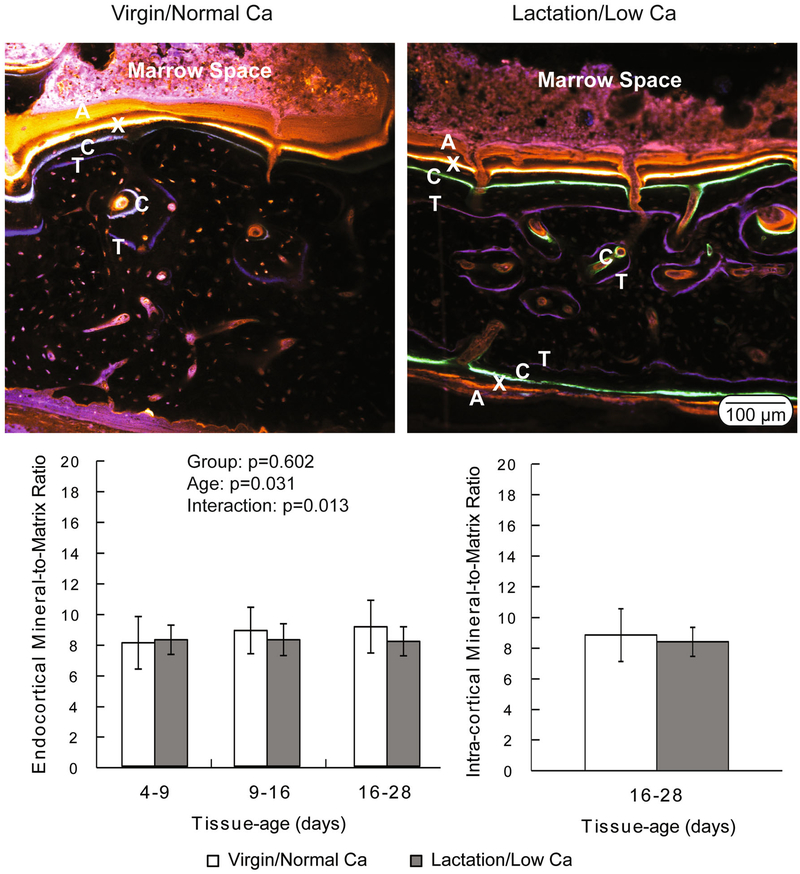
Fig. 5.
(Top) Representative confocal microscopy images demonstrating the presence of fluorochrome labels in the endocortical and intra-cortical compartments of both virgin/normal Ca and lactation/ low-Ca groups. The virgin/normal Ca controls lacked fluorochrome labelsin the periosteal compartment. Oxytetracycline (labeled ‘T’ in the images) was given on day 2; calcein (‘C’) was given on day 14; xylenol orange (‘X’) was given on day 21; and alizarin (‘A’) was given on day 26 of the recovery phase. (Bottom) Degree of mineralization (mineral-to-matrix ratio) plotted as a function of tissue age and compartment. Data are presented as the means and standard deviations. There was not a significant difference between groups in the intra-cortical compartment. Endocortically, the two-way ANOVA indicated a significant tissue age effect (p = 0.031) and a significant interaction term (p = 0.013), with no significant group effect (p =0.602)
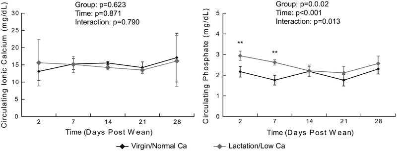
Fig. 6.
Circulating calcium and phosphate concentration measured longitudinally during the recovery phase (means and standard deviations, n = 4–5 per group per time point). Results from the repeated measures ANOVA are presented in the legend


