Abstract
Case summary
A 5-month-old cat was evaluated for a 3 week history of cough, nasal discharge, decreased appetite and weight loss. Musculoskeletal examination was normal and serum creatine kinase (CK) activity was within the reference interval. The cat was treated during the next 10 months for chronic, persistent pneumonia. Weakness then became apparent, the cat developed dysphagia and was euthanized. Post-mortem evaluation revealed chronic aspiration pneumonia and muscular dystrophy associated with beta (β)-sarcoglycan deficiency.
Relevance and novel information
This is the first report of a cat with muscular dystrophy presenting for chronic pneumonia without obvious megaesophagus, dysphagia or prominent neuromuscular signs until late in the course of the disease. The absence of gait abnormalities, marked muscle atrophy or hypertrophy and normal serum CK activity delayed the diagnosis in this cat with β-sarcoglycan deficiency.
Keywords: Muscular dystrophy, amyotrophy, dysphagia, sarcoglycan, bronchopneumonia
Introduction
Muscular dystrophies (MDs) are a heterogenous group of inherited, degenerative muscle disorders characterized by progressive muscle weakness, muscle atrophy or hypertrophy and abnormal gait.1 Dystrophin-deficient MD is the most common form of MD diagnosed in people, dogs and cats.1–4 Other forms of MD previously described in cats include laminin alpha (α)-2 deficiency and one recent report of reduced beta-sarcoglycan (β-SG) in a domestic shorthair cat.1–7 This case report describes a second cat with β-SG-deficient MD, which exhibited neuromuscular signs only late in the course of disease. This cat was initially evaluated for chronic pneumonia that was minimally responsive to treatment with relatively unremarkable gait and clinical examination, and normal serum creatine kinase (CK) activity, making the diagnosis of MD challenging.
Case description
A 5-month-old male domestic longhair cat was presented to the Veterinary Teaching Hospital at the University of Saskatchewan with a 3 week history of a raspy cough, a wet-sounding purr, serous nasal discharge, decreased appetite and weight loss. No dysphagia was reported. The cat had been routinely vaccinated and dewormed.
A male littermate of this cat had been euthanized 1 week earlier for progressive respiratory signs not responsive to antibiotics. The littermate had a 2 month history of cough, wheezing, hoarse voice, poor body condition and a short history of dysphagia. Thoracic radiographs showed a caudodorsal bronchial pulmonary pattern. Laryngeal examination and bronchial endoscopy were normal. Hematologic and biochemical evaluation were normal, including CK activity (239 U/l; reference interval [RI] 75–471 IU/l). The cat continued to deteriorate and was humanely euthanized. Post-mortem examination and histopathology results revealed multifocal-to-coalescing lesions consistent with subacute fibrinosuppurative bronchopneumonia, but no significant respiratory pathogens were isolated. The skeletal muscles were not grossly abnormal but were not evaluated histologically.
Physical examination of the cat reported here revealed a small stature and poor body condition (body condition score 3/9). Thoracic auscultation was unremarkable, but there was an intermittent bilateral serous nasal discharge and a productive cough was easily elicited on tracheal palpation. The cat was active and alert with normal gait, strength and mobility, although the owner reported that it was ‘knock kneed’ at home.
Thoracic radiographs showed a mild bronchial pattern in both caudal lung lobes, as well as a mild increase in pulmonary opacity in the cranioventral thorax consistent with an interstitial pattern. Additionally, there was questionable mild, focal thickening of the parietal pleura in the cranioventral thorax (Figure 1). Feline leukemia virus antigen and feline immunodeficiency virus antibody tests were negative. Complete blood count (CBC) and serum biochemistries were unremarkable apart from a slightly decreased creatinine concentration (40 µmol/l; RI 78–178 µmol/l) and a very mild increase in serum CK activity (574 U/l; RI 75 – 471 U/l).
Figure 1.
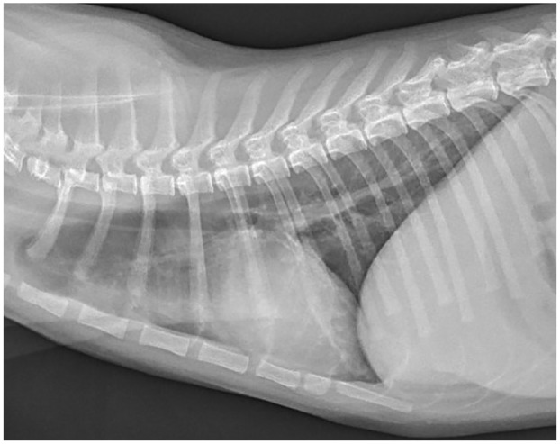
Plain lateral thoracic radiograph of the cat showing a mild bronchial pattern in the caudal lung lobes, as well as a mild increase in pulmonary opacity in the cranioventral thorax
The cat was discharged without treatment while waiting for the necropsy results of the sibling. Seven days after initial presentation (day 7), the cat experienced worsening of the cough and was re-examined. Although the owner reported the cat was still very active and playful at home, subtle weakness was noted in the pelvic limbs, attributed to the cat’s poor body condition and diminished muscle mass; therefore, no neurological examination was performed at this time. Mild crackles were detected on auscultation of the cranial ventral lung fields and pharyngeal examination revealed generalized erythema and multiple small papules. The tongue was normal. Submandibular lymph nodes were slightly enlarged.
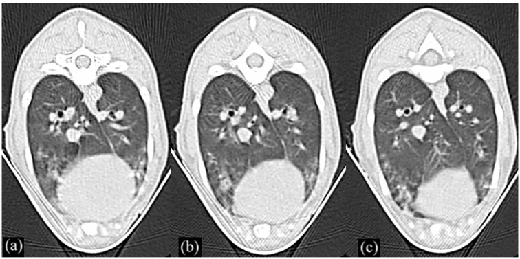
Repeat thoracic radiographs indicated a progressive interstitial pulmonary pattern, which now coalesced into a focal alveolar pattern immediately cranial to the cardiac silhouette. A same-day thoracic ultrasound identified a discrete region of atelectasis or pulmonary infiltrate/consolidation, approximately 1 cm in length and up to 5 mm in depth, in the caudal ventral aspect of the left cranial lung lobe. Several small, poorly defined pleural irregularities with prominent B-lines were identified in the remainder of the cranial and middle lung lobes. No mediastinal masses or pleural effusion were identified. A brief two-dimensional examination of the heart found no abnormalities. A thoracic CT (1 mm slice thickness) revealed patchy ground-glass opacity, greatest in the ventral lung fields, with marked peribronchial thickening and mild dilation of the peripheral ventral airways (Figure 2). Mild pleural thickening was also noted in the ventral lung margins. The overall appearance was considered most supportive of chronic inflammatory disease (pneumonia/pneumonitis). An endotracheal brushing was performed. A bronchoscopy was not attempted owing to the small stature of the patient and financial constraints.
Figure 2.
CT of the thorax. (a–c) Patchy ground-glass opacity of the lungs greatest in the ventral fields, with marked peribronchial thickening and mild dilation of the peripheral ventral airways
The critical condition of the cat on recovery from the anesthesia necessitated a short hospitalization with oxygen therapy. While recovering, the cat started to have a better appetite. No dysphagia was noted at this time. Cytologic evaluation of the sample revealed septic suppurative inflammation and culture and PCR were positive for Mycoplasma species, Neisseria species and Pasteurella multocida. Treatment was initiated with enrofloxacin (6 mg/kg PO q24h) and amoxicillin clavulanate (15 mg/kg PO q12h). Coupage and nebulization with saline or salbutamol were implemented and continued indefinitely. The owner initiated elevation of food bowls and passive range-of-motion exercises to support the mild general weakness that was noted at that time.
The cat was re-examined on day 50, at which time the owner reported that the cough was unchanged but appetite, energy level and strength were all dramatically improved. The cat continued to be in poor body and muscle condition and had developed a mild plantigrade stance, which were deemed to be associated with the chronic disease. Thoracic radiographs were unchanged and antimicrobial treatments were continued. On day 75, the cat was presented for lethargy and a more severe cough. CBC showed neutropenia (1.290 × 10⁹/l; RI 2.100–15.000 × 10⁹/l) and thoracic radiographs indicated worsening of the ventrally distributed interstitial to alveolar pattern. Doxycycline treatment was initiated (5 mg/kg PO q12h) and the other antibiotics were stopped. Five days later when the cat deteriorated, pradofloxacin (15 mg/kg PO q24h) and N-acetylcysteine (20 mg/kg PO q12h)8 were added to the doxycycline, based on suspicion of a primary ciliary dyskinesia.
On day 147, at approximately 9 months of age, the cat had clinically improved in terms of cough, appetite and strength, but thoracic radiographs showed progression of the bronchopneumonia, with an increased alveolar component noted vs the previous studies. Ciliary dyskinesia was suspected, so the cat was anesthetized and neutered, and the testicles submitted for electron microscopy. Although no overt ciliary abnormalities were identified, electron microscopy alone could not allow exclusion of ciliary dyskinesia. Despite supportive treatment and ongoing antibiotic treatment, the cat continued to deteriorate. It was only around day 225, when the cat was fostered by one of the authors (ML), that dysphagia became evident, and generalized muscle weakness more pronounced. A short-strided gait, inability to jump and capability of walking only short distances were also identified at that time. The owner declined re-evaluation, preventing further neurological evaluation and work-up for dysphagia (videofluoroscopic swallowing study, electromyography, etc).
The cat deteriorated and was humanely euthanized. Necropsy revealed poor body condition with generalized muscle atrophy and minimal body fat stores. Histopathologic evaluation of the lungs was consistent with chronic mild-to-moderate, multifocal granulomatous pneumonia with intrahistiocytic and extracellular foreign material suggesting aspiration.
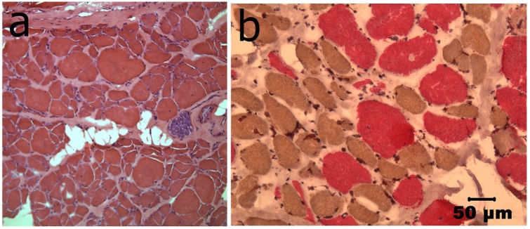
Histopathology of multiple cryosections of the vastus lateralis, biceps femoris, temporalis, esophageal, intercostal and diaphragmatic muscles showed a marked variability in myofiber size, with type 1 fiber predominance, moderate endomysial fibrosis, occasional fibers containing internal nuclei, multifocal fatty infiltration and minimal-to-mild myonecrosis consistent with a congenital myopathy or congenital muscular dystrophy (Figure 3).
Figure 3.
(a) Representative hematoxylin and eosin-stained cryosection from the diaphragm, showing excessive variability in myofiber size, moderate endomysial fibrosis, occasional myofibers showing internal nuclei and multifocal areas of mild adipose tissue infiltration. (b) Representative staining of the vastus lateralis muscle showing type 1 fiber predominance using monoclonal antibodies against slow myosin (type 1 fibers, brown stain) and fast myosin (type 2 fibers, pink stain). Bar in (b) = 50 µm for (a) and (b)
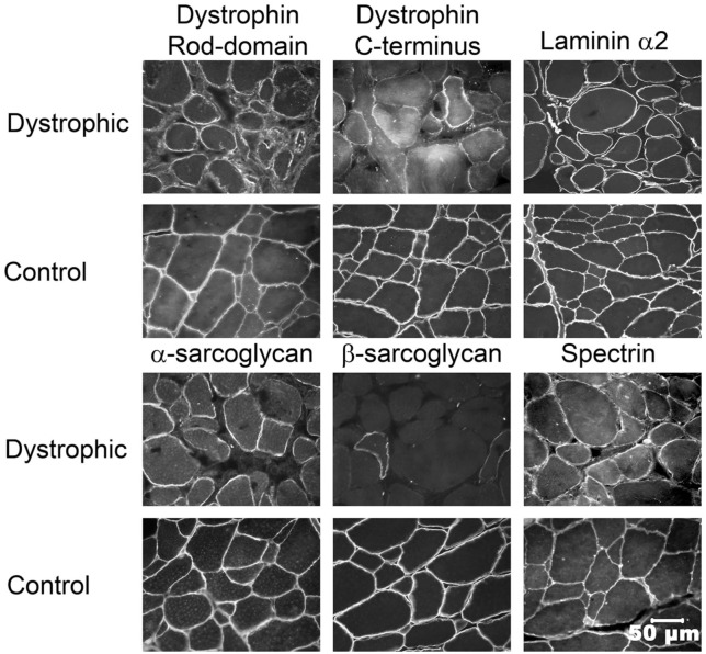
Immunofluorescence staining of cryosections cut at 9–10 µm from the vastus lateralis muscle was performed with monoclonal antibodies against the rod domain (1:20, NCL DYS1) and C-terminus (1:20, NCL DYS2) of dystrophin, β-SG (1:50, NCL-b-SARC) and spectrin (1:100, NCL-SPEC2) as a control for membrane integrity. All monoclonal antibodies were from Novocastra Laboratories, Newcastle-Upon-Tyne. Polyclonal antibodies were used against α-SG (1:1000, a gift of Dr Eva Engvall)9 and laminin α2 (1:1000, a gift of Dr Eva Engvall).10 Fluorescent labels included fluorescein isothiocyanate (FITC)-conjugated goat anti-mouse IgG (1:200, 111-095-003; Jackson ImmunoResearch) and FITC-conjugated goat anti-rabbit IgG (115-095-003; Jackson ImmunoResearch). After washing, slides were sealed with anti-fade solution (H-1500; Vector Laboratories) then observed directly with a fluorescence microscope. Markedly reduced staining for β-SG (Figure 4) with normal staining for the rod domain and C-terminus of dystrophin, α-SG and laminin α2 was present.
Figure 4.
Immunofluorescence staining of cryosections from the vastus lateralis muscle using monoclonal or polyclonal antibodies against the rod domain and C-terminus of dystrophin, spectrin, laminin alpha (α)2, and α- and beta (β)-sarcoglycan (SG). Stainings for both the rod domain and C-terminus of dystrophin, laminin α2 and α-SG were similar to control muscle while staining for β-SG was markedly reduced. Spectrin staining confirmed membrane integrity. Bar in lower right image = 50 µm for all images
To confirm the results of immunofluorescence staining, immunoblotting was performed by standard methods using extracts from the vastus lateralis muscle and archived control tissue. Primary antibodies included α-SG (1:2000), β-SG (1:2000) and gamma (γ)-SG (1:1000), all from Novocastra Laboratories. A monoclonal antibody against β-actin (1:2000, A2066; Sigma) was used as a loading control. Protein bands were detected using Super Signal West Dura Extended Duration Substrate (Thermo Scientific). Deficiency of β-SG and normal levels of α- and γ-SG was confirmed by immunoblotting (Figure 5).
Figure 5.
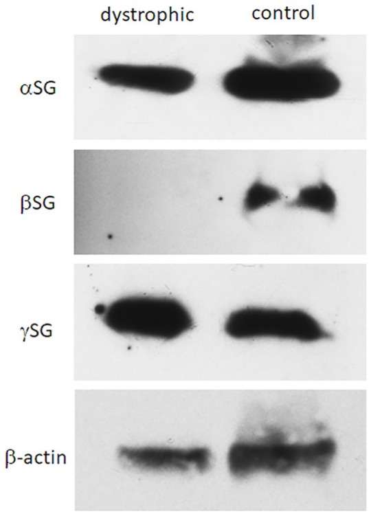
Immunoblot analysis of the vastus lateralis muscle from the affected cat and archived control cat muscle. Consistent with the immunofluorescence results, staining for beta (β)-sarcoglycan (SG) was markedly decreased or absent, while staining for alpha (α)- and gamma (γ)-SG was similar to control. Staining with a monoclonal antibody against β-actin was used as a loading control
Discussion
This is, to our knowledge, only the second description of MD associated with β-SG deficiency in a cat.6 In contrast to the cat of this report that presented for a history of chronic respiratory disease, the 6-month-old cat previously reported with partial β-SG deficiency was evaluated for a 4 month history of severe progressive muscle weakness without apparent muscle atrophy, reluctance to move, hyporeflexia and a 2 month history of respiratory distress.6 Pathological changes in the muscles evaluated were similar between both cases with marked variability in myofiber size, type 1 fiber predominance and moderate endomysial fibrosis. Reduced or absent β-SG (1:50, NCL-b-SARC) was confirmed in both cases, by immunostaining and immunoblotting. All reported dogs and the previously reported cat with sarcoglycanopathy have had moderate-to-marked increases in aspartate aminotransferasee, alanine aminotransferase and CK activity, unlike the cat reported here.2,11,12 In Duchenne muscular dystrophy, CK activity decreased in association with muscle loss and low physical activity due to disease progression.13
Mutational analysis was not performed in either the cat reported here or its deceased littermate. Although a definitive diagnosis was not reached for the littermate, we strongly suspect that this cat was suffering from the same disorder, perhaps making inheritance of the condition from asymptomatic carriers more likely than a spontaneous mutation.
Mutations in the SG complex are responsible for a subset of the human autosomal recessive muscular dystrophies known as the limb girdle muscular dystrophies (LGMDs).2,14 SGs are sarcolemmal transmembrane glycoproteins that assemble together to form the SG complex. The SG complex is an important part of the dystrophin glycoprotein complex, which links the muscle fiber actin in the cytoskeleton to the extracellular matrix, protecting the sarcolemma from tension during muscle contraction. SG deficiency has been previously reported in several dogs and one cat.6,11,12,14
MDs are uncommon in cats, with most reported cases associated with dystrophin deficiency and presenting for clinical signs of muscular weakness, a stiff, stilted or bunny-hopping gait, hypertrophy of the neck and shoulder muscles and tongue enlargement in cats less than 6 months of age.2,4,15–17
Dystrophin-deficient MD (DDMD) is caused by mutations in the very large dystrophin gene and is characterized by absence of or reduction in the dystrophin protein at the sarcolemma.1,2 The gene for dystrophin is located on the X chromosome, so reported cases are nearly always male.2,4 The hypertrophic form of DDMD is most often described in cats, causing muscular weakness. Multifocal calcified lingual nodules, megaesophagus, dilated cardiomyopathy, diaphragmatic thickening, respiratory distress and hepatosplenomegaly have all been described.2,4,15–18 Serum CK activity is markedly increased in cats with DDMD as early as a few days of age, as a lack of myofiber membrane stability leads to myofiber necrosis.1,2,4,15–17
Laminin α2 (merosin)-deficient MD has been reported in three young (2.5 to 6-month-old) female cats with generalized severe muscle atrophy and weakness, hypotonia, hyporeflexia and limited jaw mobility.1–3,5 Progressive muscle spasticity and limb contractures developed in one cat.1,3,5 Serum CK activities were moderately increased in all affected cats. Laminin α2 links dystrophin to the extracellular matrix and contributes to the stability of the muscle basement membrane. Immunofluorescence staining (Figure 4) ruled out both dystrophin and laminin α2-deficient muscular dystrophies in our cat.
Muscular dystrophy has been reported to cause respiratory signs in humans, dogs and cats.1–4,6,7,11,12,19,20 Severe muscular weakness can lead to hypoventilation, respiratory insufficiency or, occasionally, to an inability to abduct the arytenoids during inspiration or to close the glottis during eating.4,7 Enlargement of the base of the tongue in cats with hypertrophic MD causes pharyngeal and esophageal dysfunction, drooling, dysphagia and regurgitation increasing the risk of aspiration pneumonia.1,2
The unusual thing about the cat reported here was that initially the respiratory signs predominated without obvious appendicular muscle weakness. Acute respiratory insufficiency, megaesophagus and aspiration without obvious skeletal muscle weakness was previously reported in one cat with DDMD.4 Respiratory failure is a leading cause of morbidity and mortality in human patients with all forms of MD, particularly the sarcoglycanopathies, but generalized muscle weakness is usually evident.19 Impaired respiratory muscle strength has been demonstrated in humans with LGMDs and suspected in dogs with SG deficiencies.11,12,14,19,20 Respiratory expiratory muscle failure increases the risk for pulmonary sepsis, as patients cannot cough effectively and respiratory airway clearance is impaired.19 Rarely, human patients with SG deficiencies present with severe respiratory insufficiency before exhibiting remarkable appendicular weakness or disability.20
Bacterial pneumonia is a relatively rare condition in cats and is usually secondary to hematogenous spread, although inhalation and local extension from other processes (ie, pyothorax) have also been described.21,22 There was no evidence of retroviral immunosuppressive disease or systemic infection in the cat reported here, and the histopathologic findings made inhalation and impaired clearance the most likely cause of bacterial pneumonia in this case. Although there was no evidence of megaesophagus, the cat was having difficulty swallowing during the last month of life, at which point generalized neuromuscular weakness was apparent. Pharyngeal muscle weakness, dysphagia and impaired respiratory airway clearance associated with MD are considered to be the most likely cause of recurrent and persistent bronchopneumonia in this cat. Ideally, a full work-up for dysphagia and neuromuscular weakness should have been performed, including neurological examination, repeat oral and laryngeal examination under general anesthesia, and contrast video-fluoroscopy motion studies.
In addition, although the cat’s condition was improving while receiving antimicrobial treatment, it is important to note that not all cases of aspiration pneumonia need antimicrobial therapy.23 Indeed, the clinical disease might not be associated with bacterial infection but rather secondary to chemical irritation from aspirated content.23 When treating bacterial pneumonia, the current recommendation is to treat for 4–6 weeks, although shorter courses may be sufficient (10–14 days).23 In this case, decisions to extend treatment were based on clinical, hematological and radiographic findings. The positive clinical response of the cat to antimicrobial treatment lead to prolonged antimicrobial therapy, which seemed appropriate based on suspicion of an underlying incurable disease (ciliary dyskinesia). However, in the light of the final diagnosis, extended antimicrobial treatments were unlikely to be necessary.
Conclusions
Inherited neuromuscular diseases should be considered as a possibility when a young cat has recurrent or persistent bacterial pneumonia, even without obvious megaesophagus. Although most cats with MD will have marked muscle atrophy or hypertrophy and a dramatic increase in serum CK activity, that is not always the case in cats with β-SG deficiency.
Footnotes
Accepted: 13 May 2019
Conflict of interest: The authors declared no potential conflicts of interest with respect to the research, authorship, and/or publication of this article.
Funding: The authors received no financial support for the research, authorship, and/or publication of this article.
Ethical approval: This work involved the use of client-owned animal(s) only, and followed established internationally recognised high standards (‘best practice’) of individual veterinary clinical patient care. Ethical approval from a committee was not therefore needed.
Informed consent: Informed consent (either verbal or written) was obtained from the owner or legal guardian of all animal(s) described in this work for the procedure(s) undertaken. For any animals or humans individually identifiable within this publication, informed consent for their use in the publication (verbal or written) was obtained from the people involved.
ORCID iD: Juliette Bouillon  https://orcid.org/0000-0003-2926-4926
References
- 1. Shelton GD, Engvall E. Muscular dystrophies and other inherited myopathies. Vet Clin North Am Small Anim Pract 2002; 32: 103–124. [DOI] [PubMed] [Google Scholar]
- 2. Shelton GD, Engvall E. Canine and feline models of human inherited muscle diseases. Neuromuscul Disord 2005; 15: 127–138. [DOI] [PubMed] [Google Scholar]
- 3. O’Brien DP, Johnson GC, Liu LA, et al. Laminin α2 (merosin)-deficient muscular dystrophy and demyelinating neuropathy in two cats. J Neurol Sci 2001; 189: 37–43. [DOI] [PubMed] [Google Scholar]
- 4. Gambino AN, Mouser PJ, Shelton GD, et al. Emergent presentation of a cat with dystrophin-deficient muscular dystrophy. J Am Anim Hosp Assoc 2014; 50: 130–135. [DOI] [PubMed] [Google Scholar]
- 5. Poncelet L, Resibois A, Engvall E, et al. Laminin alpha2 deficiency-associated muscular dystrophy in a Maine coon cat. J Small Anim Pract 2003; 44: 550–552. [DOI] [PubMed] [Google Scholar]
- 6. Salvadori C, Vattemi G, Lombardo R, et al. Muscular dystrophy with reduced β-sarcoglycan in a cat. J Comp Pathol 2009; 140: 278–282. [DOI] [PubMed] [Google Scholar]
- 7. Martin PT, Shelton GD, Dickinson PJ, et al. Muscular dystrophy associated with α-dystroglycan deficiency in Sphynx and Devon Rex cats. Neuromuscul Disord 2008; 18: 942–952. [DOI] [PMC free article] [PubMed] [Google Scholar]
- 8. Buur JL, Diniz PPVP, Roderick KV, et al. Pharmacokinetics of N-acetylcysteine after oral and intravenous administration to healthy cats. Am J Vet Res 2013; 74: 290–293. [DOI] [PubMed] [Google Scholar]
- 9. Liu LA, Engvall E. Sarcoglycan isoforms in skeletal muscle. J Biol Chem 1999; 274: 38171–38176. [DOI] [PubMed] [Google Scholar]
- 10. Leivo I, Engvall E. Merosin, a protein specific for basement membranes of Schwann cells, striated muscle, and trophoblast, is expressed late in nerve and muscle development. Proc Natl Acad Sci U S A 1988; 85: 1544–1548. [DOI] [PMC free article] [PubMed] [Google Scholar]
- 11. Munday JS, Shelton GD, Willox S, et al. Muscular dystrophy due to a sarcoglycan deficiency in a female Dobermann dog. J Small Anim Pract 2015; 56: 414–416. [DOI] [PubMed] [Google Scholar]
- 12. Deitz K, Morrison JA, Kline K, et al. Sarcoglycan-deficient muscular dystrophy in a Boston terrier. J Vet Intern Med 2008; 22: 476–480. [DOI] [PubMed] [Google Scholar]
- 13. Manzur AY, Kinali M, Muntoni F. Update on the management of Duchenne muscular dystrophy. Arch Dis Child 2008; 93: 986–990. [DOI] [PubMed] [Google Scholar]
- 14. Schatzberg S, Whittemore J, Morgan E, et al. Sarcoglynopathy in 3 dogs. Abstract number 88, Proceedings of the American College of Veterinary Internal Medicine 21st Annual Veterinary Medicine Forum; 2003, June 4-7, Charlotte, NC. [Google Scholar]
- 15. Carpenter JL, Hoffman EP, Romanul FC, et al. Feline muscular dystrophy with dystrophin deficiency. Am J Pathol 1989; 135: 909–919. [PMC free article] [PubMed] [Google Scholar]
- 16. Winand NJ, Edwards M, Pradhan D, et al. Deletion of the dystrophin muscle promoter in feline muscular dystrophy. Neuromuscul Disord 1994; 4: 433–445. [DOI] [PubMed] [Google Scholar]
- 17. Gaschen F, Burgunder J-M. Changes of skeletal muscle in young dystrophin-deficient cats: a morphological and morphometric study. Acta Neuropathol (Berl) 2001; 101: 591–600. [DOI] [PubMed] [Google Scholar]
- 18. Gaschen L, Lang J, Lin S, et al. Cardiomyopathy in dystrophin-deficient hypertrophic feline muscular dystrophy. J Vet Intern Med 1999; 13: 346–356. [DOI] [PubMed] [Google Scholar]
- 19. Fayssoil A, Ogna A, Chaffaut C, et al. Natural history of cardiac and respiratory involvement, prognosis and predictive factors for long-term survival in adult patients with limb girdle muscular dystrophies type 2C and 2D. PLoS One 2016; 11: e0153095. [DOI] [PMC free article] [PubMed] [Google Scholar]
- 20. Walter M, Dekomien G, Schlotter-Weigel B, et al. Respiratory insufficiency as a presenting symptom of LGMD2D in adulthood. Acta Myol 2004; 23: 1–5. [PubMed] [Google Scholar]
- 21. Foster SF, Martin P. Lower respiratory tract infections in cats: reaching beyond empirical therapy. J Feline Med Surg 2011; 13: 313–332. [DOI] [PMC free article] [PubMed] [Google Scholar]
- 22. Macdonald ES, Norris CR, Berghaus RB, et al. Clinicopathologic and radiographic features and etiologic agents in cats with histologically confirmed infectious pneumonia: 39 cases (1991–2000). J Am Vet Med Assoc 2003; 223: 1142–1150. [DOI] [PubMed] [Google Scholar]
- 23. Lappin MR, Blondeau J, Boothe D, et al. Antimicrobial use guidelines for treatment of respiratory tract disease in dogs and cats: Antimicrobial Guidelines Working Group of the International Society for Companion Animal Infectious Diseases. J Vet Intern Med 2017; 31: 279–294. [DOI] [PMC free article] [PubMed] [Google Scholar]


