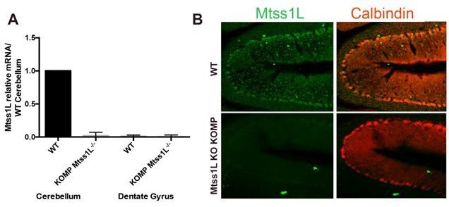
Author response image 2. Validation of Mtss1L-/- KO KOMP mice.

(A) RT-qPCR data confirmed Mtss1L deletion in Mtss1L-/- KO KOMP mice in the cerebellum. Notably Mtss1L mRNA levels in the dentate gyrus were undetectable in wildtype (WT) and Mtss1L-/- KO KOMP mice. mRNA was isolated from cerebellum and dentate gyrus tissue from 6 weeks old (WT) and Mtss1L-/- KO KOMP mice. RT-qPCR data are from three animals per genotype, with each sample run in duplicate (p < 0.05, t-test). (B) Mtss1L immunohistochemistry was performed on WT and Mtss1L-/- KO KOMP cerebellum. Purkinje cells were labeled with anti-Calbindin. Although quality of anti-Mtss1L is very low as seen in top panels, Mtss1L expression pattern in the cerebellum of WT mice overlapped with Calbindin+ Purkinje cells. Mtss1L expression was absent in the cerebellum of Mtss1L KO KOMP mice validating both the mouse line and the antibody.
