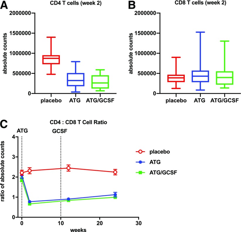
Figure 3.
CD4:CD8 T-cell ratio declined with low-dose ATG and ATG/GCSF treatment. Absolute counts were generated at the 2-week time point by multiplying lymphocyte complete blood cell counts with flow cytometry–detected CD4+ (A) or CD8+ (B) T-cell percentages within the lymphocyte gate. Significant differences in CD4+ cells were identified between each of the treatment arms and placebo (P < 0.001 for both pairwise comparisons). No significant differences were observed for CD8+ cells. Longitudinal CD4:CD8 T-cell ratios (C) were determined using frequencies in the lymphocyte gate. Average measures with SDs by treatment arm are shown. Dotted lines in C denote initiation of ATG and last dose of GCSF. The CD4:CD8 T-cell ratios were significantly different at the postbaseline time points for those treated with ATG or ATG/GCSF vs. placebo (all P < 0.001).

