Abstract
Purpose:
Our study aims at evaluating the efficacy and safety of botulinum toxin A in the early treatment of sixth nerve palsy in type 2 diabetic patients.
Methods:
This study is a prospective and interventional clinical case series of patients presenting with acute onset of sixth cranial nerve palsy, who received injection botulinum toxin A.
Results:
Thirty-one cases were included in the study. 58% of the study subjects had incomplete palsy at presentation (abduction deficit -1 to -3) and 42% had complete palsy (-4 and -5). The median dosage of injection was 5 U (range 3--6 U). The median follow-up period is 2 months. The P value shows that there is statistically significant improvement in head turn, ocular deviation in primary position, and improvement in abduction between baseline and 1 week (P-value <0.001), 1 month (P-value <0.001) and 2 month (P-value <0.001) postinjection follow-up visits. 90.3% of patients had full resolution of symptoms in the last follow-up visit. 83.9% of patients were successfully treated.
Conclusion:
Early injection of botulinum toxin A in select patients with acquired sixth nerve palsy due to diabetes is a safe and efficient treatment option in alleviating symptoms, restoring function and quality of life and reducing need for surgical interventions in future.
Keywords: Botulinum toxin A, diabetic mononeuropathy, sixth cranial nerve palsy
One of the causes for an isolated sixth cranial nerve palsy is diabetic mononeuroppathy.[1] Acute onset of diplopia causes considerable morbidity and loss of productivity due to inability to work and perform other day-to-day activities. Although most cases resolve spontaneously, it often takes 3--6 months for complete resolution of symptoms.[2] Injection of botulinum toxin A (botox) which is derived from clostridium botulinum into the antagonist medial rectus prevents contracture and allows for faster recovery of the paralyzed lateral rectus muscle[3,4] Despite a long history of use of botulinum toxin in ocular muscle paralysis, very few reports are published on its efficacy and safety in diabetic mononeuropathy. Hence, we report the efficacy and safety of botulinum toxin A in the early treatment of sixth nerve palsy in type 2 diabetic patients.
Methods
This is a prospective and interventional clinical case series of patients presenting with acute onset of sixth cranial nerve palsy and who received injection of botulinum toxin A between August 2015 and February 2018. This study was conducted at a tertiary eye care hospital in Coimbatore, South India. This study was approved by the Institutional Ethics Committee.
Patients with acquired sixth cranial nerve palsy due to diabetes, with no other neurological defects and who received injection botulinum toxin A were included in the study. Informed consent was taken from all patients prior to receiving the injection.
All patients underwent a complete ophthalmological evaluation including best corrected uniocular visual acuity and refraction, ocular motility, measurement of ocular deviation at distance and near, by prism bar cover test (PBCT), anterior segment, and fundus evaluation.
Head posture was measured using goniometer with the patient fixing at distance target (6 m). Diplopia charting was performed using red--green Armstrong goggles and a vertical streak light source held at 50 cm. Abduction deficit was noted on the scale described by Scott and Kraft: zero (normal), -1 (to 75% full rotation), -2 (to 50% full rotation), -3 (to 25% full rotation), -4 (to midline), and -5 (inability to abduct to the midline).[5] Abduction deficit of -1 and -2 were grouped as incomplete palsy and an abduction deficit of -3 and above as complete palsy.[6] The angle of deviation was measured in prism diopters by prism cover test in primary position with normal eye fixing target at 6 m (primary deviation). The dosage of injection of botulinum toxin A and its immediate adverse drug effects if any were also noted.
Head posture, abduction limitation, and ocular deviation in primary position with the normal eye fixing for distance were recorded at presentation, and at follow-up visits 7, 30, and 60 days post-injection botulinum toxin A. Success was defined as a primary deviation of less than 10 prism diopters for distance, subjective improvement in diplopia-free field and 2 score improvement in abduction, at the 60 days follow-up visit.
The patients who presented with uncontrolled diabetes mellitus were asked to consult a diabetologist for control of blood sugar levels prior to giving the injection. A fasting sugar level was measured on the day of injection, and the patients received the injection only if FBS <150 mg/dl.
Method of reconstitution of botulinum toxin
We had used commercially available botulinum toxin agent, BOTOX (Allergan Inc., CA) in the study. Each vial of botox contains 50 units of botulinum toxin A which is reconstituted gently with 1 ml sterile nonpreserved saline, so that the reconstituted injection yields 5 units per 0.1 ml. The reconstituted drug was used immediately, and the remaining preserved in the lower compartment of a refrigerator for further use within 4 days.
Method of administration of botox
All injections were given by a single experienced strabismologist under local subtenon (lignocaine) or topical anesthesia (proparacaine), the topical eye drops administered every 5 min for 4 times prior to injection. Pulse oximeter was connected and patients pulse rate closely monitored throughout the procedure. The surgical field was cleaned and draped under sterile aseptic precautions. The treatment dosage was based on the primary deviation and limitation of abduction, with dose ranging from 2.5 to 6 U at one time in one muscle. (Based on deviation - For vertical muscles, and for horizontal strabismus of less than 20 prism diopters: 1.25 Units to 2.5 Units in any 1 muscle For horizontal strabismus of 20 prism diopters to 50 prism diopters: 2.5 Units to 5 Units in any 1 muscle- Information given in the guidelines inset enclosed in the vial. We have modified these guidelines by injecting upto 6 units in a muscle) After applying the eyelid speculum, the medial rectus muscle was grasped with a rectus holding forceps firmly at its insertion and the eye is held in primary position. A 27 gauge needle on a 1 cc disposable syringe having the required amount of drug was inserted into the medial rectus muscle belly about 5 mm away from its insertion without electromyographic guidance, the needle being parallel to the medial orbital wall and the drug is slowly injected. After injecting, the needle is slowly withdrawn after 10--15 s to minimize drug extravasation and spread. The absence of ballooning of conjunctiva or tenons tissue after injection signifies drug entry into the muscle belly and not into the subconjunctival or subtenons space. The patient is advised to apply topical antibiotics four times a day for a week and review after a week.
Statistical analysis
Wilcoxon sign rank test was used to find out the significant difference between pre and postinjection visits. P value less than 0.05 considered as statistically significant.
Results
A total of 31 cases were included in the study, 77.4% of the study subjects being male and 22.6% female. Mean (SD) age of the study population is 54.7 (11.29) years and it ranges from 30 years to 80 years. The mean duration between the onset of diplopia and intervention with botulinum toxin injection was 26 days (range 3--60). 58% of the study subjects had incomplete palsy at presentation (abduction deficit -1 to -3) and 42% had complete palsy (-4 and -5). The median dosage of injection was 5 U (range 3--6 U) [Table 1].
Table 1.
Dosage of Botox Injection
| Botox dose | n | % |
|---|---|---|
| 2.5-3 | 2 | 6.5 |
| 3.1-4 | 4 | 12.9 |
| 4.1-5 | 24 | 77.4 |
| 5.1-6 | 1 | 3.2 |
| Total | 31 | 100 |
In this study, the median preinjection head turn was 20° (mean 19.52) improved to 10° (mean 7.42) 1 week postinjection (P < 0.001) and was nil by 1 month postinjection (mean 1.83) maintained at 2 month (Mean -1.00) follow-up visit (P < 0.001) [Table 2].
Table 2.
Comparison of Head posture, Primary deviation and abduction limitation -pre and post-botox Injection
| Baseline | 1 week | 1 month | 2 month | |
|---|---|---|---|---|
| Head posture | ||||
| Mean in degree (SD) | 19.52 (7.78) | 7.42 (6.44) | 1.83 (5.65) | 1.00 (4.03) |
| IQR | 15.0-20.0 | 0.0-10.0 | 0.0 to 0.0 | 0.0 to 0.0 |
| P | - | <0.001 | <0.001 | <0.001 |
| Primary deviation | ||||
| Mean deviation in PD (SD) | 29.26 (8.64) | 11.73 (7.90) | 2.87 (10.51) | 0.03 (7.52) |
| IQR | 20.0-35.0 | 6.0-16.0 | 0.0 to 8.0 | 0.0 to 0.0 |
| P | - | <0.001 | <0.001 | <0.001 |
| Abduction limitation | ||||
| Mean (SD) | -3.32 (0.75) | -2.19 (1.01) | -0.94 (1.18) | -0.29 (0.82) |
| IQR | -4.0 to -3.0 | -3.0--1.0 | 1.0 to 0.0 | 0.0 to 0.0 |
| P | - | <0.001 | <0.001 | <0.001 |
The median ocular deviation at baseline was 30 prism diopters (mean - 29.26) improved to 12 (mean - 7.42) 1 week postinjection (P < 0.001) and to 0 (mean -2.87) by 1 month postinjection, the improvement maintained at 2 month postinjection follow-up [Table 2 and Fig. 1].
Figure 1.
Pre (a-c) and post (6 month) Botulinum Toxin A injection (d-f) in a 60 year old lady with left lateral rectus palsy showing resolution of esotropia and improvement in abduction
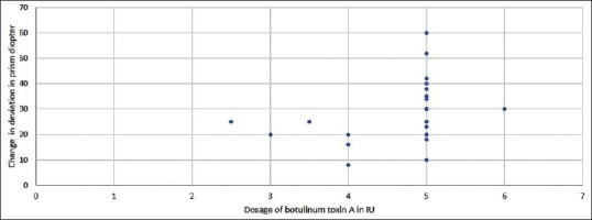
We have grouped the patients into two groups based on the botox dosage as 2.5--4 U (group 1) and 4.1--6 U (group 2). The patients in group 1 (19.53% of total) showed a mean change in deviation of 19 PD (8--25) at 2 months postinjection. Group 2 patients (80.64%) showed a mean change in deviation of 31.68 PD (10--60) at 2 months postinjection. (Deleted the conclusion that response was dose dependent) [Fig. 2].
Figure 2.

Distribution on the change in primary deviation in prism diopter with respect to the dosage of Botulinum toxin A
The median restriction to abduction was -3 (mean - 3.32) at baseline, which improved to -2 (mean - 2.19) at 1 week postinjection (P < 0.001), -1 (mean - 0.94) at 1 month postinjection (P < 0.001) and 0 (full abduction) (mean- 0.29) 2 month postinjection (P < 0.001) [Table 2]. The P value shows that there is a significant improvement in head turn between baseline and 1 week (P value <0.001), 1 month (P value < 0.001) and 2 months (P-value <0.001). Similarly, P value also shows a statistically significant reduction in ocular deviation in primary position and improvement in abduction between the baseline (preinjection) values and each of the postinjection follow-up visits. 35.5% of patients had no diplopia in straight gaze 1 week postinjection, which increased to 67.7% at 1 month post and 87.1% at 2 months postinjection, respectively.
The median follow-up period is 2 months (IQR 2--12 months). 90.3% of patients had full resolution of symptoms in the last follow-up visit. 45.16% of the patients (n = 14) had follow-up period of ≥4 months. Mean residual deviation was 0 in this group. None of them had a recurrence of esotropia.
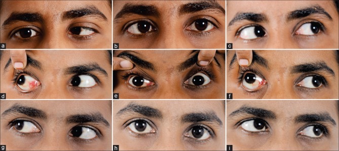
At 1 week post-botox injection, we observed ptosis with adduction limitation in six patients (19.35%) and limitation of adduction alone in 1 patient (3.2%.). These complications resolved at 1 month follow-up visit [Fig. 3]. We did not have any case of vertical rectus palsy.
Figure 3.
32 year old lady with right lateral rectus palsy and -4 abduction limitation (a-c), 4 days postinjection had ptosis, exotropia and adduction limitation (-4) (d-f). With complete resolution of all symptoms at 1 month (g-i)
According to our defined criteria of success (deviation – <=10 prism D, No diplopia and 2 score improvement in abduction at 60th day follow-up), in 83.9% of patients the treatment was successful [Table 2, study data].
Discussion
Botulinum toxin, an exotoxin produced by Clostridium botulinum causes flaccid paralysis of skeletal muscle by blocking acetylcholine release at the neuromuscular junction.[4,7]
The treatment of acute sixth nerve paresis due to diabetic mononeuropathy with botulinum toxin A injection is controversial due to high incidence of spontaneous improvement with time. Most report suggest spontaneous recovery by 3--6 months of time.[6]
A sub-sect of these patients who are intolerant to diplopia and suffering from inability to perform routine and work related activities during this waiting period are the ones who would most be benefitted by botulinum toxin A injection.[8] Prisms though another option helps only in alleviating symptoms in straight gaze and is not very effective in treating the incomitance in the lateral gaze.
Subtenons space injection of botulinum toxin A has also been described,[9,10] the advantages being stated by authors as simpler and lesser time for procedure, no need for EMG guidance[10,11] and no major side effects.[12] In our study, the injection was given intramuscularly under local/topical anesthesia and the procedure took less than 5 min. The side effects observed were transient ptosis and adduction limitation and resolved completely within 1 month of injection. We did not see the complications of postinjection subconjunctival hemorrhage or globe perforation in any of our patients. Ptosis and acquired vertical deviations are the reported commonest complications encountered with botulinum toxin A and vision-threatening complications are rare.[12] Also repeated use of botulinum toxin A injection is safe.
Other studies have also reported lower complication rate and that the treatment can be given in an office setting. It was also found to delay more definitive incisional surgical intervention.[6]
Botulinum toxin A injection helps patients to achieve significant increase in diplopia free field within a week of injection and hence greatly reduces morbidity and time off from work and professional activities.[12] It tends to achieve better outcome in terms of ocular alignment.[3] By paralyzing the antagonist medial rectus muscle, it also hastens recovery of function of paralyzed lateral rectus muscle[3] It is an accepted preoperative therapy that may alleviate medial rectus restriction. Botox injection may be curative in a certain percentage of patients with lateral rectus palsy.[13] This procedure is proved to be simple, safe, cheap, effective, and avoids the risks of general anesthesia.[3]
Hence, we believe that early injection of botulinum toxin A, within 2--3 weeks of onset of nerve palsy would give the best results due to early rehabilitation and preventing contracture of antagonist medial rectus muscle.
The limitations of our study are that we could not compare out outcomes with the cohort of patients managed conservatively, who refused to receive injection botulinum toxin A.
Conclusion
Early injection of botulinum toxin A in select patients with acquired sixth nerve palsy due to diabetes, with FBS <150 mg%, who complain of troublesome double vision and hence inability to perform day-to-day and professional activities, is a safe and efficient treatment option in alleviating symptoms, and restoring function and quality of life.
Financial support and sponsorship
Nil.
Conflicts of interest
There are no conflicts of interest.
References
- 1.Patel SV, Mutyala S, Leske DA, Hodge DO. Incidence, associations, and evaluation of sixth nerve palsy using a population-based method. Ophthalmology. 2004;111:369–75. doi: 10.1016/j.ophtha.2003.05.024. [DOI] [PubMed] [Google Scholar]
- 2.King AJ, Stacey E, Stephenson G, Trimble RB. Spontaneous recovery rates for unilateral sixth nerve palsies. Eye (Lond) 1995;9:476–8. doi: 10.1038/eye.1995.110. [DOI] [PubMed] [Google Scholar]
- 3.Quah BL, Ling YL, Cheong PY, Balakrishnan V. A review of 5 years’ experience in the use of botulinium toxin A in the treatment of sixth cranial nerve palsy at the Singapore National Eye Centre. Singapore Med J. 1999;40:405–9. [PubMed] [Google Scholar]
- 4.Repka MX, Lam GC, Morrison NA. The efficacy of botulinum neurotoxin A for the treatment of complete and partially recovered chronic sixth nerve palsy. J Pediatr Ophthalmol Strabismus. 1994;31:79–83. doi: 10.3928/0191-3913-19940301-04. [DOI] [PubMed] [Google Scholar]
- 5.Scott AB, Kraft SP. Botulinum toxin injection in the management of lateral rectus paresis. Ophthalmology. 1985;92:676–83. doi: 10.1016/s0161-6420(85)33982-9. [DOI] [PubMed] [Google Scholar]
- 6.Holmes JM, Beck RW, Kip KE, Droste PJ, Leske DA. Botulinum toxin treatment versus conservative management in acute traumatic sixth nerve palsy or paresis. J AAPOS. 2000;4:145–9. [PubMed] [Google Scholar]
- 7.Crouch ER. Use of botulinum toxin in strabismus. Curr Opin Ophthalmol. 2006;17:435–40. doi: 10.1097/01.icu.0000243018.97627.4c. [DOI] [PubMed] [Google Scholar]
- 8.Broniarczyk-Loba A, Czupryniak L, Nowakowskans O. Botulinum toxin A in the early treatment of sixth nerve palsy-induced diplopia in type 2 diabetes. Diabetes Care. 2004;27:846–7. doi: 10.2337/diacare.27.3.846. [DOI] [PubMed] [Google Scholar]
- 9.Chen YH, Sun MH, Hsueh PY, Kao LY. Botulinum injection for the treatment of acute esotropia resulting from complete acute abducens nerve palsy. Taiwan J Ophthalmol. 2012;4:140–3. [Google Scholar]
- 10.Kao LY, Chao AN. Subtenon injection of botulinum toxin for the treatment of traumatic sixth nerve palsy. J Pediatrc Ophthalmology Strabismus. 2003;40:27–30. doi: 10.3928/0191-3913-20030101-09. [DOI] [PubMed] [Google Scholar]
- 11.Sanjari MS, Falavarjani KG, Kashkouli MB, Aghai GH, Nojomi M, Rostami H. Botulinum toxin injection with and without electromyographic assistance for treatment of abducens nerve palsy: A pilot study. J AAPOS. 2008;12:259–62. doi: 10.1016/j.jaapos.2007.11.006. [DOI] [PubMed] [Google Scholar]
- 12.Kowal L, Wong E, Yahalom C. Botulinum toxin in the treatment of strabismus. A review of its use and effects. Disabil Rehabil. 2007;29:1823–31. doi: 10.1080/09638280701568189. [DOI] [PubMed] [Google Scholar]
- 13.Wu X. Botulinum toxin A in treatment of the sixth cranial nerve palsy. Zhonghua Yan Ke Za Zhi. 2002;38:457–61. Chinese. [PubMed] [Google Scholar]


