Left ventricular outflow tract (LVOT) obstruction is a common devastating complication of transcatheter mitral valve replacement (TMVR), with few treatment options, including alcohol septal ablation, causing myocardial and conduction tissue damage, or cardiac surgery.
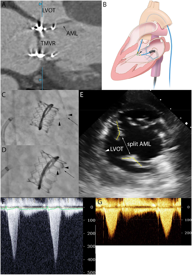
A 63-year-old male with previous mitral annuloplasty (30mm Physio 2 ring; Edwards Lifesciences) developed severe LVOT obstruction after transseptal TMVR-in-ring with a 26 Sapien3 valve (Edwards Lifesciences). Transthoracic echocardiography and cardiac CT demonstrated systolic anterior motion (SAM) of a 23mm long anterior mitral valve leaflet (AML) (Figure 1A). The LVOT gradient was 120mmHg despite a neo-LVOT area of 600mm2. He was NYHA class III despite medical therapy and developed hemolytic anemia. The patient and heart team agreed to modify the prophylactic LAMPOON procedure1 and lacerate the AML as rescue therapy.
Figure: The “rescue” LAMPOON procedure.
[A] Cardiac-gated CT shows SAM after TMVR. [B] Illustration of concept: a kinked guidewire lacerates the AML in the direction of the black arrow. [C] Fluoroscopy in RAO with kinked mid-shaft of the Astato guidewire (black arrow) sheathed in two microcatheters (black arrow heads) guided by a transseptal sheath and transfemoral catheter. [D] The guidewire is electrified and pulled to lacerate the native AML. [E] Mid-gastric transesophageal view confirming a split AML parted away from the LVOT. [F-G] Severe LVOT gradient is treated.
We used a novel simplified approach to lacerate the AML from tip-to-base with an electrified guidewire (Figure 1B). The Sapien3 valve frame provided a barrier to deleterious advancement of the lacerating edge into the aortomitral curtain and aortic root. An 0.014” guidewire (Astato XS 20, Asahi) was focally kinked and denuded to form a lacerating edge. The guidewire was insulated at both ends by microcatheters (Finecross, Terumo) and positioned at the AML centerline, between an SL-1 transseptal sheath and transfemoral JL3.5 guiding catheter (Figure 1C). The guidewire was pulled and electrified with three 5-second applications of 70W radiofrequency energy and 5% dextrose flush until adjacent to the Sapien3 valve frame (Figure 1D and Video 1). Leaflet splitting was confirmed on echocardiography (Figure 1E) before LAMPOON system retrieval. LVOT gradient reduced to 20mmHg (Figure 1F–G). The patient was discharged home the next day and, on follow-up, was NYHA class I and able to cycle uphill, with laminar flow in the LVOT and no significant gradient.
VIDEO LEGEND.
Transthoracic echocardiogram and cardiac-gated CT images show SAM after TMVR. Fluoroscopy and transesophageal echocardiography shows the kinked guidewire between the transseptal sheath and transfemoral guiding catheter being retracted during radiofrequency energy application. The guidewire can be seen advancing towards the Sapien3 valve frame and microbubbles are seen from guidewire electrification. Transesophageal echocardiogram confirms midline laceration of the anterior mitral leaflet in front of the LVOT.
This case highlights two important points. First, the length and mobility of the AML must be considered at pre-procedural assessment, as this may cause LVOT obstruction despite a capacious neo-LVOT. Second, LAMPOON is feasible after TMVR as a treatment for LVOT obstruction caused by SAM. Unlike preventative LAMPOON, the procedure is simplified but only the protruding leaflet tip is lacerated, so the efficacy of leaflet separation needs to be further evaluated.
Acknowledgments
Funding:
Supported by Z01-HL006040 and Z01-HL006039 from the Division of Intramural Research, NHLBI, NIH
ABBREVIATIONS
- TMVR
Transcatheter Mitral Valve Replacement
- LAMPOON
Laceration of the Anterior Mitral leaflet to Prevent Outflow ObstructioN
- LVOT
Left Ventricular Outflow Tract
- SAM
Systolic Anterior Motion
- NYHA
New York Heart Association
- AML
Anterior Mitral valve Leaflet
Footnotes
Publisher's Disclaimer: This is a PDF file of an unedited manuscript that has been accepted for publication. As a service to our customers we are providing this early version of the manuscript. The manuscript will undergo copyediting, typesetting, and review of the resulting proof before it is published in its final citable form. Please note that during the production process errors may be discovered which could affect the content, and all legal disclaimers that apply to the journal pertain.
Disclosures:
DHS is a proctor/advisory for Edwards Lifesciences, Medtronic, and Boston Scientific.
Remaining authors have no disclosures.
REFERENCE
- 1.Babaliaros VC, Greenbaum AB, Khan JM, Rogers T, Wang DD, Eng MH, O’Neill WW, Paone G, Thourani VH, Lerakis S, Kim DW, Chen MY and Lederman RJ. Intentional Percutaneous Laceration of the Anterior Mitral Leaflet to Prevent Outflow Obstruction During Transcatheter Mitral Valve Replacement: First-in-Human Experience. JACC Cardiovasc Interv. 2017;10:798–809. [DOI] [PMC free article] [PubMed] [Google Scholar]


