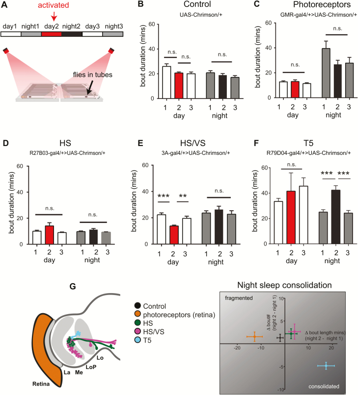
Figure 4.
Activation of T5 motion-detection neurons consolidates nighttime sleep. (A) Flies expressing a red-light-activated channelrhodopsin (Chrimson) were placed in the recording set up and sleep was analyzed under baseline conditions on day 1 (normal white light from 8 am to 8 pm, followed by 12 hours darkness at night), an activated condition on day 2 (normal white light + 12 hours red light illumination from 8 am to 8 pm, followed by 12 hours darkness at night), and recovery conditions (same as baseline). Analyses in this figure are from the same dataset as in Figure 3. (B–F) Sleep bout duration during the day and night across all three conditions for the UAS-Chrimson/+ control (B) and red-light-activated optic lobe circuits (C–F). (G) Summary graph showing sleep consolidation for the aforementioned strains, viewed as a change in sleep bout number/duration following red-light activation (night 2), normalized to baseline sleep conditions (night 1). Activation of T5 neurons (cyan) led to consolidation of nighttime sleep as observed by an increase in sleep bout duration and decrease in sleep bout number. n = 31 flies in (B), 33 flies in (C), 33 flies in (D), 30 flies in (E), and 47 flies in (F). Error bars indicate the SEM. **p < 0.01, ***p < 0.001, one-way analysis of variance with Tukey’s multiple comparisons.

