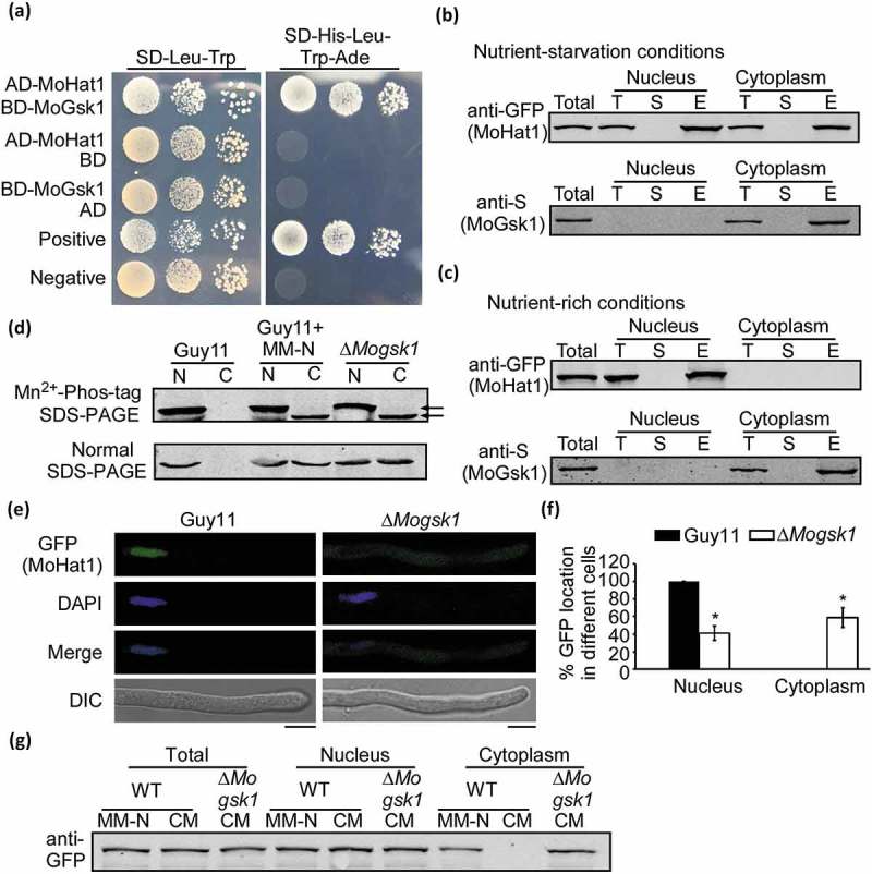
Figure 10.

Phosphorylation of MoHat1 by MoGsk1 is important for its nuclear localization. (a) Yeast two-hybrid analysis for the interaction of MoHat1 and MoGsk1. Plasmids pGBKT7-Lam and pGADT7-T were used as a negative control. (b and c) Co-IP analysis for the interaction of MoHat1-MoGsk1. Nucleus and cytoplasm proteins were extracted separately using the Nuclear and Cytoplasmic Protein Extraction Kit under nutrient-rich and starvation conditions and incubated with anti-GFP or anti-S agarose and then eluted for western blot detecting using anti-S or anti-GFP antibodies. (d) Nucleus and cytoplasm proteins were extracted separately and the phosphorylation level of MoHat1 in the ∆Mogsk1 mutant was analyzed by Mn2+-Phos-tag SDS-PAGE and normal SDS-PAGE, respectively. (e) Localization of MoHat1 in the ∆Mogsk1 mutant was observed by confocal fluorescence microscope (Zeiss LSM710, 63x oil). Scale bars: 5 μm. (f) Quantification of GFP fluorescence localized in the nucleus and the cytoplasm in wild-type Guy11 and the ∆Mogsk1 mutant. Asterisks represent significant differences (Duncan’s new multiple range test, p < 0.01). (g) Western blot analysis of MoHat1 in Guy11 (WT) and ∆Mogsk1 mutant strains. Total, nuclear and cytoplasmic proteins were extracted separately and detected with the GFP antibody. ‘CM’ indicates the nutrient-rich conditions and ‘MM-N’ indicates nutrient starvation conditions.
