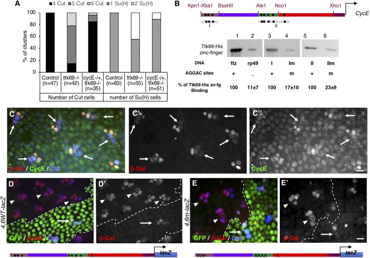
Figure 4.
CycE expression is transcriptionally repressed by Ttk69. (A) The integrity of a Ttk-mutant SO was rescued under CycE heterozygous conditions. Histogram showing the percentage of SO harboring four (black bars) or more (gray bars). Cut-positive cells (left), and one (hatched bars) or two (white bars) Su(H)-positive cells (right), located outside (control) and inside Ttk69 clones in CycE+/+ or CycEAR95/+ heterozygous backgrounds. (B) DNA-mediated Ttk69-His-zinc finger pull-down assay. (Top) Diagram of Ttk69-binding sites (black dots) in the CycE promoter. (Bottom) Magnetic beads were coated with: lane 1, ftz promoter bearing AGGAC-binding sites (positive control); lane 2, rp49 an AGGAC-free promoter (negative control); lanes 3 and 5, I and II regions of the CycE promoter, respectively; and lanes 4 and 6, Im and IIm regions of the CycE promoter, respectively, in which AGGAC-binding sites were replaced by an unrelated ACTGC sequence. (C) The 4.6WT fragment recapitulates endogenous CycE expression in adult bristle sensory cells. Immunostaining of 24-hr APF pupae. Sensory cells in blue (outer cells indicated with arrows). β-Gal in red (shown as a separate channel in the middle C’), CycE in green (shown as a separate channel in C") immunoreactivity. (D and E). Expression pattern of the 4,6WT-lacZ (D and D’) and 4,6m-lacZ (E and E’) CycE transcriptional reporters in control (arrows) and Ttk69-mutant SOs (arrowheads). Ttk69 clones outlined by a white line were detected by the absence of GFP (green). Sensory cells (Cut immunoreactivity, blue) and expression of CycE transcriptional reporters (β-Gal immunoreactivity, red). Bar, 10 μm for (C and D). APF, after pupal formation; β-Gal, β-galactosidase; SO, sensory organ.

