Abstract
Hydroxyethylsulfonate (isethionate (Ise)) present in mammalian tissues is thought to be derived from aminoethylsulfonate (taurine), as a byproduct of taurine nitrogen assimilation by certain anaerobic bacteria inhabiting the taurine-rich mammalian gut. In previously studied pathways occurring in environmental bacteria, isethionate is generated by the enzyme sulfoacetaldehyde reductase IsfD, belonging to the short-chain dehydrogenase/reductase (SDR) family. An unrelated sulfoacetaldehyde reductase SarD, belonging to the metal-dependent alcohol dehydrogenase superfamily (M-ADH), was recently discovered in the human gut sulfite-reducing bacterium Bilophila wadsworthia (BwSarD). Here we report the structural and biochemical characterization of a sulfoacetaldehyde reductase from the human gut fermenting bacterium Bifidobacterium kashiwanohense (BkTauF). BkTauF belongs to the M-ADH family, but is distantly related to BwSarD (28% sequence identity). The crystal structures of BkTauF in the apo form and in a binary complex with NAD+ were determined at 1.9 and 3.0 Å resolution, respectively. Mutagenesis studies were carried out to investigate the involvement of active site residues in binding the sulfonate substrate. Our studies demonstrate the presence of sulfoacetaldehyde reductase in Bifidobacteria, with a possible role in isethionate production as a byproduct of taurine nitrogen assimilation.
Keywords: NADH/NADPH, Nitrogen assimilation, Organosulfur degradation, Sulfoacetaldehyde reductase, X-ray crystallography
Introduction
Isethionate (Ise) is ubiquitous in the environment, and is also present in mammalian tissues [1]. There is no known pathway for isethionate biosynthesis in animals. Instead, it is thought to originate from metabolism of bile salt-derived taurine by anaerobic gut bacteria [1,2]. Both taurine and isethionate serve as substrates for sulfate- and sulfite-reducing bacteria (SSRB) in the gut, which use the sulfonate-derived sulfite as a terminal electron acceptor (TEA) for anaerobic respiration, producing toxic H2S [3,4]. Relatively few taurine-reducing SSRB have been isolated, including the prominent human gut SSRB Bilophila wadsworthia [4]. In contrast, isethionate-reducing SSRB appear to be common, and include several Desulfovibrio and Desulfitobacterium species closely related to gut SSRB [3,5].
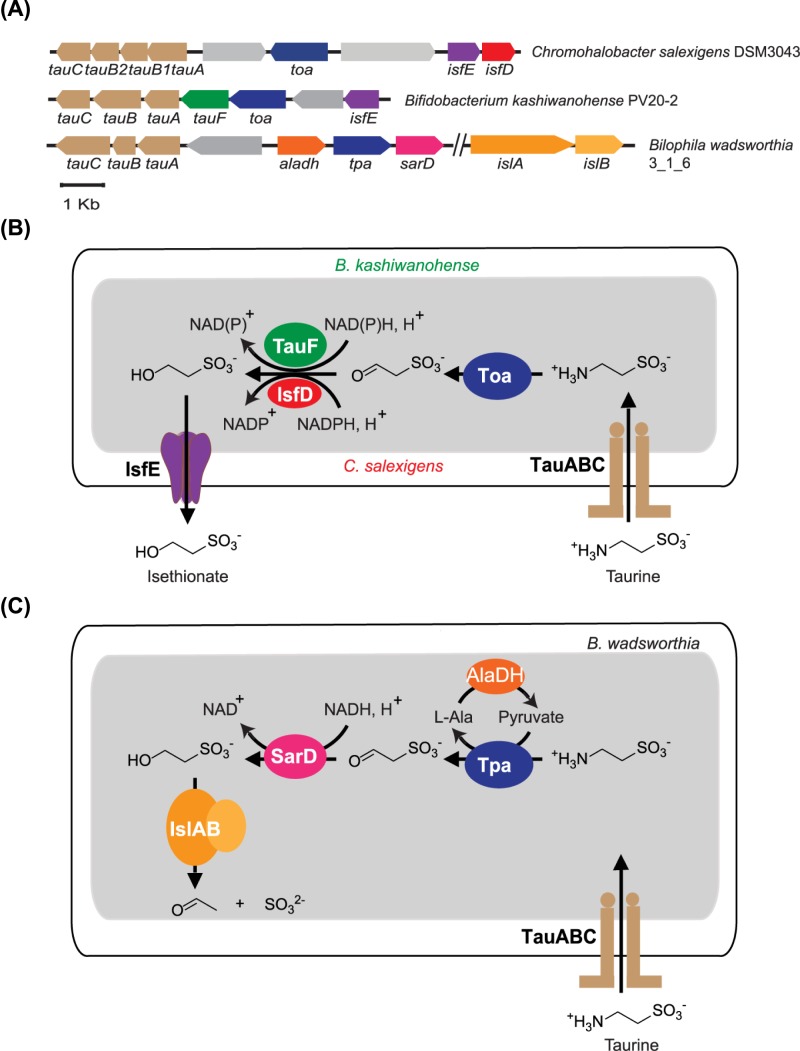
Conversion of taurine into isethionate has been observed in a mixed culture of anaerobic gut bacteria [1]. Although the specific gut bacterial strains catalyzing this reaction have not been isolated to date, the pathway for isethionate production as a byproduct of taurine nitrogen assimilation has been studied in detail in certain environmental bacteria, including Klebsiella oxytoca TauN1 and Chromohalobacter salexigens DSM3043, and involves enzymes and transporters arranged in gene clusters (Figure 1A) [6,7]. In this pathway, taurine is imported by a taurine ABC transporter (TauABC), and converted into sulfoacetaldehyde by taurine:oxoglutarate aminotransferase (Toa), generating glutamate as an intermediate for nitrogen metabolism. The NADPH-dependent sulfoacetaldehyde reductase (IsfD), belonging to the SDR family, generates isethionate as a waste produce, which is exported by the putative isethionate exporter (IsfE) (Figure 1B).
Figure 1. Gene clusters and metabolic pathways involving different sulfoacetaldehyde reductases isozymes.
(A) Gene clusters containing the sulfoacetaldehyde reductases IsfD (in Chromohalobacter salexigens), TauF (in Bifidobacterium kashiwanohense) and SarD (in Bilophila wadsworthia). (B) Schematic diagram showing two bacterial taurine nitrogen assimilation pathways relying on different sulfoacetaldehyde reductase isozymes. The pathway in C. salexigens relies on IsfD (red), while the putative pathway in B. kashiwanohense relies on TauF (green). (C) Schematic diagram showing the taurine dissimilation pathway in B. wadsworthia.
We recently reported the crystal structure of IsfD from K. oxytoca TauN1, in complex with NADPH and isethionate [8], and showed that it is part of a group of 3-hydroxyacid dehydrogenases, which are ubiquitous in bacteria. However, IsfD-containing gene clusters for isethionate formation have only been predicted in bacteria belonging to the phylum Proteobacteria [6,8], and not in strict anaerobic bacterial taxa that are abundant in the gut. Apart from IsfD, a new sulfoacetaldehyde reductase (SarD), belonging to the metal-dependent alcohol dehydrogenase superfamily (M-ADH) family, was recently reported in B. wadsworthia (BwSarD) [9,10]. This enzyme is involved in a pathway for taurine dissimilation, in which isethionate is generated as an intermediate, and further degraded to acetate and H2S instead of being secreted (Figure 1C).
While examining the occurrence of taurine-related genes in different human gut bacteria, we noticed the occurrence of a putative gene cluster for taurine nitrogen assimilation, present in B. kashiwanohense PV20-2, an isolate from human infant faeces (Figure 1A) [11]. The gene cluster resembles those previously reported by Cook et al. [6]. However, instead of IsfD, it contains a member of the M-ADH family, with only 28% identicality to the previously reported BwSarD. Here we report the biochemical and structural characterization of this M-ADH enzyme, which we refer to as BkTauF. We demonstrate that it is a novel sulfoacetaldehyde reductase, which differs from BwSarD in active site residues and metabolic function.
Materials and methods
General materials and methods
Lysogeny broth (LB) medium was purchased from Oxoid Limited (Hampshire, U.K.). Methanol and acetonitrile used for liquid chromatography-mass spectrometry (LC-MS) were high-purity solvents from Concord Technology (Minnesota, U.S.A.). Water used in this work was ultrapure deionized water from Millipore Direct-Q. Chemicals were purchased from Sigma–Aldrich, Geneview, J&K, Amresco or Solarbio. Oligonucleotide primers were synthesized by Tsingke Biological Technology (Beijing, China). DNA plasmid Mini prep, fragment gel extraction and restriction endonucleases clean up kits were from Tiangen (Beijing, China). Talon Cobalt resins were purchased from Clonetech (California, U.S.A.). All protein purification chromatographic experiments were performed on an ‘ÄKTA pure’ or ‘ÄKTA prime plus’ FPLC machine equipped with appropriate columns (GE Healthcare). Protein concentrations were determined according to the method of Bradford [12] using bovine serum albumin (BSA) as a standard, or using NanoDrop One (Thermo Fisher Scientific). The extinction coefficient for each protein was obtained using the ExPASy ProtParam tool.
Gene syntheses and cloning
The codon-optimized gene fragment of BkTauF (UniProt accession: A0A0A7I0A5) was synthesized by General Biosystems (Anhui, China), and inserted into pET-28a-HMT at the SspI restriction site. The resulting plasmid HMT-BkTauF contains, in tandem: a His6-tag, maltose binding protein (MBP) and a Tobacco Etch Virus (TEV) protease cleavage site, followed by BkTauF [13]. The expression of an MBP-BkTauF fusion protein allows purification with an amylose affinity column, followed by cleavage with TEV protease, resulting in high purity of BkTauF.
The original construct was resistant to TEV protease cleavage likely due to structural hindrance. A linker (GGG) between the TEV cleavage site and the N-terminus of BkTauF was thus introduced. New DNA fragment, produced by PCR using the original construct as template and primers HMT-GGG-BkTauF-F and HMT-GGG-BkTauF-R (Supplementary Table S1), was inserted into pET-28a-HMT at the SspI site by Gibson assembly [14], forming HMT-GGG-BkTauF. Recombinant plasmids were confirmed by DNA sequencing.
Expression and purification of BkTauF
The HMT-GGG-BkTauF plasmid was transformed into Escherichia coli BL21 (DE3) cells. The transformant was grown in LB medium containing kanamycin (50 μg/ml) at 37°C in flasks in a shaker incubator at 220 rpm, and induced for the expression of MBP-BkTauF with 0.3 mM isopropyl β-d-1-thiogalactopyranoside (IPTG) for 16 h at 18°C. Typically, cells from 4 l culture were harvested by centrifugation at 8000×g for 10 min and lysed with French press (Panda plus, Niro Soavi Co., Italy) at 14000 psi in 200 ml buffer A (20 mM Tris/HCl, pH 7.5, 200 mM KCl, and 5 mM β-mercaptoethanol (BME)) containing 25 µg/ml DNaseI and 1 mM phenylmethanesulfonyl fluoride (PMSF). The lysate was then centrifuged at 20000×g for 10 min at 4°C, and the cell debris was discarded.
Nucleic acid was removed by precipitation with 1% streptomycin sulfate. The protein solution was then loaded on to a 10 ml TALON column (GE Healthcare). The column was washed with buffer A, and protein was eluted with buffer A containing 150 mM imidazole. The eluate was dialyzed against buffer A for 4 h to remove imidazole, then loaded on to a column packed with 40 ml amylose resin (New England Biolabs, Massachusetts, U.S.A.). The amylose column was washed with buffer A, and the protein was eluted with buffer A containing 10 mM maltose. TEV protease was added (TEV/BkTauF 1:20 mass ratio), and the solution was dialyzed overnight against 2 l buffer A. The dialyzed sample was loaded on to a 10 ml TALON column to retain His6-MBP and His6-TEV protease. The flow-through was collected and dialyzed against 2 l of buffer B (20 mM Tris/HCl, pH 8.0, 5 mM BME) for 4 h before it was loaded on to a 10 ml Q sepharose high performance column (GE Healthcare). The column was eluted with a linear salt gradient from 100 to 500 mM KCl. A prominent peak containing BkTauF was collected and concentrated to a final volume of 4 ml (5 mg/ml) using a centrifugal concentrator (30 K MWCO; Millipore). This protein solution was then injected to a Superdex200 gel filtration column and eluted with buffer C (20 mM Tris pH 7.5, 200 mM KCl, 1 mM dithiothreitol (DTT)). The eluate from gel filtration column was re-concentrated and the buffer exchanged by repeated concentration and dilution with storage buffer (10 mM HEPES, pH 7.4, 50 mM KCl, 1 mM TCEP (tris (2-carboxyethyl)phosphine)). The concentrations of purified BkTauF were calculated from its absorption at 280 nm (ԑ280 nm = 22920 M−1cm−1), measured using a NanoDrop One. The final protein concentration is approximately 10 mg/ml. The purified protein was examined on a 10% SDS polyacrylamide gel.
Determination of the oligomeric state of BkTauF
A 5 mg/ml solution of BkTauF was analyzed by gel filtration as described above, with a 4 ml injection volume and elution with buffer C at 2 ml min–1. A solution of molecular weight markers was analyzed under the same conditions. Thyroglobulin bovine (669 kDa), Horse apoferritin (443 kDa), Sweet potato β-Amylase (200 kDa), BSA (66 kDa), Bovine carbonic anhydrase (29 kDa) were used as molecular weight markers (Sigma MWGF 1000-1KT). The molecular weights of the proteins analyzed by gel filtration were calculated from their elution volume using a second-degree polynomial for the relationship between log (molecular weight) and retention time.
Crystallization, data collection and structure determination of BkTauF
Initial screening of BkTauF crystals was performed using an automated liquid handling robotic system (Gryphon, Art Robbins, California, U.S.A.) in 96-well format by the sitting-drop vapor diffusion method. The screens were set up at 295 K using various sparse matrix crystal screening kits from Hampton Research and Molecular Dimensions. Several crystallization conditions gave diamond-shaped crystal. After further optimization using the hanging-drop vapor-diffusion method in 24-well plates, we obtained crystals large enough for single crystal X-ray diffraction studies. The best condition yielding crystals was 0.2 M ammonium acetate, 0.1 M Bis-Tris, pH 5.5, 25% W/V PEG 3350. Crystals were flash-cooled in liquid nitrogen using reservoir solution containing 30% glycerol as cryo-protectant. Diffraction data were collected on BL17U1 and BL18U1 at Shanghai Synchrotron Radiation Facility (SSRF). The dataset was indexed, integrated and scaled using HKL3000 suite [15]. Molecular replacement was performed by PHENIX [16] using 1VHD as a search model [17]. The structure was manually built according to the modified experimental electron density using Win Coot [18] and further refined by PHENIX [16] in iterative cycles. Table 1 contains the statistics for data collection and final refinement. All structural figures were generated with UCSF Chimera [19]
Table 1. Data collection and refinement statistics for the BkTauF_apo (PDB ID:6JKO) and BkTauF_NAD+ crystal (PDB ID:6JKP).
| Crystals | TauF_apo | TauF_NAD+ |
|---|---|---|
| λ for data collection (Å) | 0.9795 | 0.9795 |
| Data collection | ||
| Space group | P 1 21 1 | P 1 21 1 |
| Cell dimension (Å) | ||
| a, b, c (Å) | 70.468 112.502 93.406 | 70.881 112.317 93.573 |
| α, β, γ (°) | 90 107.335 90 | 90 107.529 90 |
| Resolution | 32.3-1.9 (1.968–1.9) | 42.19-3.008 (3.115–3.008) |
| Rmerge | 0.604 (0.082) | 0.800 (0.193) |
| Average I/σ (I) | 11.78 (2.0) | 6.7 (1.2) |
| Completeness (%) | 99.50 (95.71) | 96.92(83.03) |
| Redundancy | 3.5/3.4 | 2.8/3.4 |
| Z | 4 | 4 |
| Refinement | ||
| Resolution | 32.3-1.9 Å | 42.19-3.008 Å |
| Number of reflections | 108623 | 27019 |
| Rfactor/Rfree (10% data) | 0.1566/0.1927 | 0.1998/0.2557 |
| RMSD length (Å) | 0.006 | 0.002 |
| RMSD angle (°) | 0.76 | 0.45 |
| Number of atoms | ||
| Protein | 1504 | 1504 |
| Ligands | 4 | 117 |
| Water | 1107 | 1 |
| Ramachandran plot (%) | ||
| Most favored | 98.66 | 96.26 |
| Additionally allowed | 1.34 | 3.54 |
Sulfoacetaldehyde reduction assays
The substrate sulfoacetaldehyde was introduced as a bisulfite adduct using the method reported by Denger and Cook [20]. To determine the optimal pH for sulfoacetaldehyde reduction, a 200 μl reaction solution containing one of various buffers, pH in a range of 4.0–11.0, 5 mM sulfoacetaldehyde, and 1 mM NADH, was pre-mixed in a 96 well plate, and the reaction was initiated by the addition of 0.5 μg BkTauF. The absorbance at 340 nm was monitored using a Tecan M200 plate reader with 15 s intervals for 2–3 min at room temperature (RT).
To determine the Michaelis–Menten parameters for sulfoacetaldehyde reduction, the reaction was conducted in 50 mM Tris/HCl at the optimal pH of 7.5, and the concentration of one substrate was varied (0–2.3 mM for sulfoacetaldehyde, 0–0.8 mM for NADH/NADPH), in the presence of a saturating concentration of the second substrate (0.5 mM NADH or 5 mM sulfoacetaldehyde). ΔA340nm and the extinction coefficient of NAD(P)H (6220 M−1cm−1) were used to calculate the rates of the reactions. GraphPad Prism 6.0 was used to extract the kinetic parameters.
Isethionate oxidation assays
A 200 μl reaction solution containing 50 mM as one of various buffers in a range of pH 8.0–11.5, 0.1 M isethionate and 1 mM NAD+, was pre-mixed in a 96-well plate, and the reaction was initiated by the addition of 2 μg BkTauF. The absorbance at 340 nm was monitored using a Tecan M200 plate reader with 15 s intervals for 2–3 min at RT.
To determine the Michaelis–Menten parameters for isethionate oxidation, the reaction was conducted in 50 mM 3-(cyclohexylamino)-2-hydroxy-1-propanesulfonic acid (CAPSO), at the optimal pH of 10.0, and the concentration of one substrate was varied (0–80 mM for isethionate, 0–1 mM for NAD+ or 0–5 mM for NADP+), in the presence of a saturating concentration of the second substrate (1 mM NAD+ or 100 mM isethionate).
To test the substrate specificity of BkTauF, the reaction mixture contained 50 mM CAPSO, pH 10.0, 2 μg BkTauF, 1 mM NAD+, and 0.1 M substrate (ethanol (EtOH), ethylene glycol (EG), ethanolamine (EA), 3-hydroxypropane-1-sulfonate, 3-hydroxypropionic acid (3HPA) or isethionate). To compare the activities of wild-type (WT) BkTauF and its mutants, the reaction mixture contained 50 mM CAPSO, pH 10.0, 2 μg of the WT or mutant BkTauF, 0.1 M isethionate and 1 mM NAD+.
LC-MS detection of sulfoacetaldehyde formation
The sulfoacetaldehyde product of isethionate oxidation with BkTauF was detected by derivatization with 2,4-dinitrophenylhydrazine (DNPH) (J&K). A 200 μl reaction mixture, containing 50 mM CAPSO, pH 10.0, 5 μg BkTauF, 0.1 M isethionate and 1 mM NAD+, was incubated for 10 min at 30°C. Two negative controls, omitting either BkTauF or isethionate, were also prepared. One hundred microliters of reaction solution was mixed with 1.1 ml of sodium acetate solution (0.73 M), then 800 μl DNPH (0.04%) solution was added. The mixture was incubated at 50°C for 1 h and then filtered prior to LC-MS analysis.
LC-MS analysis was performed on an Agilent 6420 Triple Quadrupole LC/MS instrument (Agilent Technologies), on an Agilent ZORBAX SB-C18 column (4.6 × 250 mm). The column was equilibrated with 75% of 0.1% formic acid in H2O, 25% of 0.1% formic acid in CH3CN, and developed at a flow rate of 1.0 ml/min from 25 to 65% CH3CN. UV detection was set at 360 nm.
Reconstitution of the metal cofactor for BkTauF
The BkTauF was dialyzed at 4°C overnight against storage buffer containing 10 mM EDTA, and then against storage buffer without EDTA. The metal cofactor was then reconstituted by incubation with a 20-fold excess of (NH4)2Fe(SO4)2.6H2O or ZnSO4 for 30 min at RT. Reconstitution with Fe2+ was carried out under an argon atmosphere [21]. The reconstituted proteins were then assayed in a reaction system containing 2 μg BkTauF, 50 mM CAPSO buffer pH 10.0, 0.1 M isethionate and 1 mM NAD+, total volume 200 μl in a 96-well plate. The absorbance at 340 nm was monitored using a Tecan M200 plate reader with 15 s intervals for 2–3 min at RT.
Site-directed mutagenesis
Three single amino acid point mutations in the enzyme active site, F252A, T257A, F265A, were introduced by site-directed mutagenesis using primers listed in Supplementary Table S1, and confirmed by sequencing. A 25 μl of PCR contained 100 ng HMT-GGG-BkTauF plasmid as template, 0.4 μM forward and reverse primers, and the Fast Alteration DNA Polymerase (KM101 from Tiangen, Beijing, China). The 17-cycle PCR mixture was digested by DpnI to remove the template before transformed into E. coli competent cells provided by the manufacturer (Tiangen). The WT and mutant BkTauF proteins were purified by Talon affinity column, followed by TEV protease cleavage. The cleaved protein was collected in the flow-through when applied to the same Talon cobalt column to retain His6-MBP and His6-TEV, and concentrated for enzyme activity assays using isethionate and NAD+ as substrates. BkTauF purified with this protocol was approximately 85% pure.
Sequence alignments
MUSCLE [22] was used to construct multiple sequence alignments. Sequence logos were plotted using WebLogo [23].
Results
BkTauF is a dimer in solution
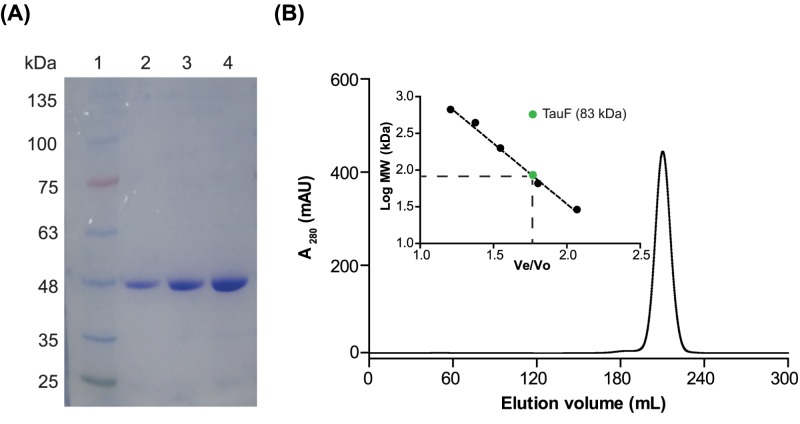
BkTauF was recombinantly produced and purified to near homogeneity through multiple chromatographic steps. The purified protein was examined on a 10% SDS/PAGE gel (Figure 2A). The gel filtration elution profile of purified BkTauF showed a single symmetric peak centered at 208.6 ml (Figure 2B). The observed molecular weight for BkTauF was 83 kDa (Figure 2B, inset), whereas the calculated molecular weight for BkTauF monomer is 41 kDa. This suggests that BkTauF exists as a dimer in solution.
Figure 2. SDS/PAGE and gel filtration analyses of BkTauF.
(A) SDS/PAGE analysis. 10% SDS gel with: lane 1, molecular weight marker; lanes 2–4, 1, 2, 4 µg of BkTauF. (B) Elution profile of BkTauF using Superdex 200 gel filtration chromatography to determine its molecular weight, estimated to be 83 kDa.
Enzyme kinetics of BkTauF
BkTauF catalyzed both sulfoacetaldehyde reduction and isethionate oxidation, as measured using a continuous spectrophotometric assay monitoring the decrease/increase in absorbance at 340 nm due to consumption/production of NAD(P)H. The optimal reaction pH for sulfoacetaldehyde reduction was 7.5, while that for isethionate oxidation was 10.0 (Supplementary Figure S1).
Enzyme kinetic parameters for the forward and reverse reactions were measured at the respective optimal pH conditions and summarized in Table 2. Although both NADH and NADPH could serve as substrates, the KM for NADH was ten-fold lower than that of NADPH (Table 2 and Supplementary Figure S2), suggesting a preference for NADH as the reductant. This is in contrast with IsfD, which uses NADPH but not NADH as a reductant [6,8].
Table 2. Enzyme kinetic parameters.
| Substrate | kcat (s−1) | KM (mM) | kcat/KM (M−1s−1) |
|---|---|---|---|
| Sulfoacetaldehyde | 22.83 ± 0.68 | 0.50 ± 0.04 | 4.61 × 10⁴ |
| Isethionate | 0.97 ± 0.03 | 4.44 ± 0.59 | 2.18 × 10² |
| NADH | 18.67 ± 0.52 | 0.06 ± 0.01 | 3.01 × 10⁵ |
| NADPH | 18.99 ± 1.13 | 0.73 ± 0.08 | 2.62 × 10⁴ |
| NAD+ | 1.30 ± 0.02 | 0.08 ± 0.00 | 1.58 × 10⁴ |
| NADP+ | 0.80 ± 0.02 | 0.77 ± 0.08 | 1.03 × 10³ |
LC-MS detection of oxidation of isethionate to sulfoacetaldehyde by BkTauF
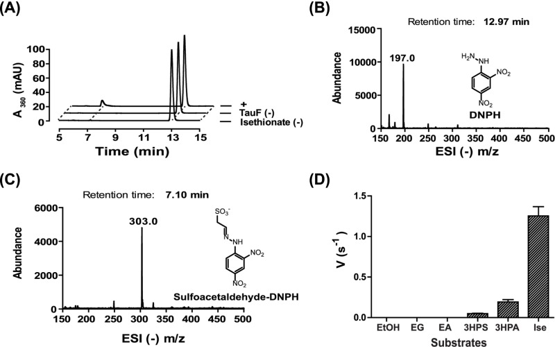
To demonstrate that sulfoacetaldehyde is the product of isethionate oxidation by BkTauF, we carried out LC-MS analysis following derivatization of the product with DNPH [8]. The LC elution profile for the assay mixture contained two major peaks with retention time 7.10 and 12.97 min corresponding to sulfoacetaldehyde-DNPH and DNPH, respectively (Figure 3A). The sulfoacetaldehyde-DNPH peak was absent from the negative controls omitting either BkTauF or isethionate. The ESI (−) (m/z) for both peaks matched with the calculated mass (Figure 3B,C).
Figure 3. LC-MS assay to detect sulfoacetaldehyde formation and spectroscopic assay to demonstrate substrate specificity.
(A) The elution profile of the LC-MS assays monitoring absorbance at 360 nm. (B) The ESI (−) m/z spectrum of the DNPH peak in A). (C) The ESI (−) m/z spectrum of the sulfoacetaldehyde-DNPH peak in (A)). (D) Spectroscopic assays monitoring absorbance at 340 nm using various alcohol substrates including EtOH, EG, EA, 3-hydroxypropane-1-sulfonate (3HPS), 3HPA and isethionate (Ise).
Substrate specificity of BkTauF
Various alcohols were tested as substrates for BkTauF. No activity was detected for EtOH, EG or EA. However, 5 and 15% of enzyme activity were detected for 3-hydroxypropane-1-sulfonate and 3HPA, respectively, possibly reflecting structural similarities to isethionate (Figure 3D).
The metal cofactor of BkTauF
Enzymes in the M-ADH family are known to utilize Zn2+ or Fe2+ as the catalytic metal [24]. Catalytic activity of BkTauF was abolished by chelation with EDTA, and recovered up to 85% by addition of Zn2+ and 15% by addition of Fe2+, suggesting that the physiological metal cofactor for BkTauF is Zn2+ (Supplementary Figure S3).
Crystal structure of BkTauF
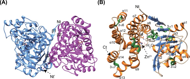
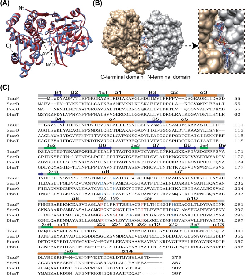
Crystal structures of BkTauF, both in the apo form and in complex with the NAD+ cofactor, were solved at 1.9 and 3.0 Å resolution, respectively. The asymmetric unit contains four monomers. The quaternary prediction server PISA suggests that BkTauF forms a dimer, consistent with its oligomeric state in solution determined by gel filtration chromatography (Figure 2B). The dimerization interface resembles that of E. coli lactaldehyde reductase FucO (PDB: 1RRM) [25], stabilized by an anti-parallel β sheet formed between the N-terminal β1 and β2 strands of the two protomers (Figure 4A). Each monomer consists of an N-terminal domain (α1–α5 and β1–β9; residues 1–178) with Rossmann fold, and a C-terminal helical domain (α6-α14; residues 179–375), similar to other structurally characterized M-ADH enzymes (Figure 4B).
Figure 4. Crystal structures of BkTauF.
(A) Quaternary structure of BkTauF. Strands β1 and β2 involved in dimer interface subunit interaction are labeled. (B) Subunit structure of BkTauF is shown in ribbon. α-helices, β strands and 310 helices are shown in orange, blue, and green, respectively.
The apo and NAD+-bound structures are nearly identical, with RMSD of 0.37 Å over 375 Cα atoms (Figure 5A). The cleft between N-terminal domain and C-terminal domain accommodates the protein active site containing NAD+ and the catalytic divalent metal ion. The electron density is well defined for NAD+ (Figure 5B), which is bound in a configuration similar to that in other M-ADH enzymes [25]. Out of the four monomers in the asymmetric unit, only one contains the catalytic Zn(II) ion, coordinated to Asp192, Gln196, His261 and His275. Structure-based sequence alignments between BkTauF, BwSarD, FucO (PDB: 1RRM) [25], and DhaT (PDB: 3BFJ) [26] show conservation in secondary structure, and metal-coordinating residues except that Gln196 is atypical in the M-ADH family, with His being more common at that position (Figure 5C).
Figure 5. Nucleotide interaction in the structure of BkTauF and structure-based sequence alignments.
(A) The apo and holo (in complex with NAD+) structures of BkTauF in red and blue, respectively, are superimposed. (B) Surface diagram show the nucleotide binding pocket in BkTauF. The N- and C-terminal domains are displayed in gray and tan, respectively. Inset: zoomed-in view with 2mFo-DFc electron density map of NAD+, displayed at 1σ. (C) Structure-based sequence alignments (Sequence identities between BkTauF, BwSarD, EcFucO and KpDhaT are 28.1, 28.1 and 30.5%, respectively). The zinc ion coordination residues are highlighted in blue, and the substrate-interacting residues are highlighted in red.
Site-directed mutagenesis
Despite attempts to soak the crystals with isethionate, we were unable to obtain a structure with the sulfonate substrate bound. Furthermore, the putative isethionate-binding site adjacent to the catalytic Zn2+ is very open, which precluded molecular docking. The present crystal structure could represent an ‘open’ conformation, and further conformational changes could occur subsequent to isethionate binding to form an enclosed active site resembling that of other M-ADH enzymes like FucO [25]. Nevertheless, we attempted to identify candidate active-site residues responsible for interaction with isethionate by examining the crystal structure.
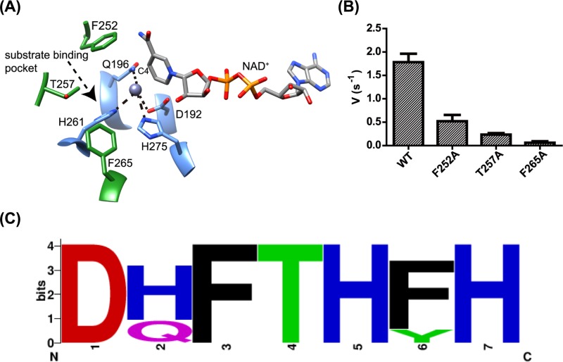
The position of isethionate is constrained by the requirements of the M-ADH catalytic mechanism, which requires coordination of the hydroxyl O-atom to Zn2+, and hydride transfer from C1 of isethionate to C4 of the NAD+. We identified Phe252, Thr257 and Phe265 surrounding the active-site cavity as potential substrate-interacting residues (Figure 6A). Alanine mutants of these three residues were assayed for isethionate oxidation, and their activities were compared with that of the WT enzyme. Activities were almost completely abolished in the T257A and F265A mutants, while the F252A mutant retained approximately 28% of the WT activity (Figure 6B). We examined the 65 BkTauF homologs in the UniRef cluster UniRef50_ A0A0J8DBY5 (where each member shares ≥50% sequence identity and ≥80% overlap with the seed sequence of the cluster [27], and observed that Phe252 and Thr257 are conserved in these proteins, while Phe265 is conservatively replaced with Tyr in a number of proteins (Figure 6C), consistent with their association with the active-site cavity in these proteins. In addition, the Zn2+ binding amino acid Gln196 is poorly conserved and replaced with the isosteric His in a number of proteins.
Figure 6. Active site structure and residues of BkTauF.
(A) Zoomed-in view of the active site of BkTauF. The substrate-interacting residues are displayed in green and labeled. Metal-coordinated residues are displayed in blue and labeled. Metal coordination is indicated by dashed lines. (B) Enzyme activities of BkTauF active site mutants. (C) Sequence logo showing the conservation of active site residues (D192, Q196, F252, T257, H261, F265 and H275) in close homologs of BkTauF in the UniRef cluster UniRef50_ A0A0J8DBY5.
Gene clusters for taurine nitrogen assimilation in anaerobic fermenting bacteria
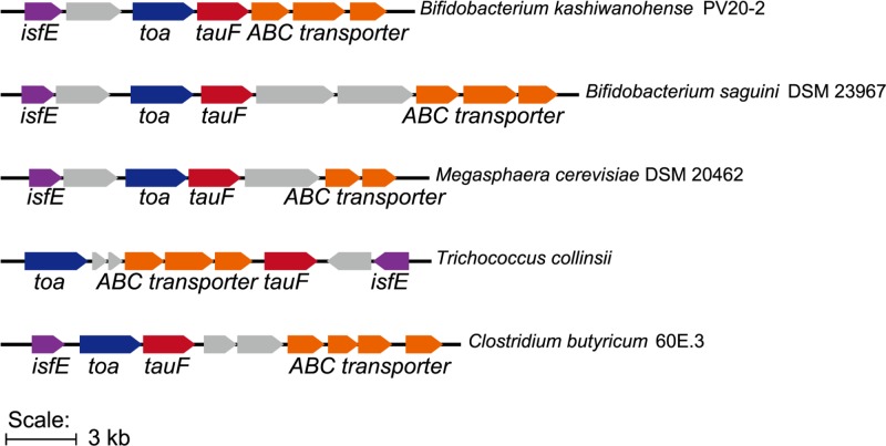
We examined the genome neighborhood of 65 BkTauF homologs in UniRef50_ A0A0J8DBY5, and observed the occurrence of TauF-containing gene clusters similar to that of B. kashiwanohense, present in several fermenting bacteria including strict anaerobes from the human gut microbiome (Figure 7). These resemble the IsfD-containing gene clusters for taurine nitrogen assimilation.
Figure 7. Gene clusters containing close homologs of BkTauF.
Many of the close homologs of BkTauF in the UniRef cluster UniRef50_ A0A0J8DBY5 are present in strict anaerobic bacteria, and associated with genes putatively involved in taurine nitrogen assimilation and isethionate secretion.
Discussion
Determination of the crystal structure of BkTauF in complex with NAD+, together with mutagenesis of active site residues, enabled us to better understand the structural requirements for substrate binding and catalysis. Interestingly, the primary sequence of BkTauF is distantly related to that of the previously reported BwSarD (only 28% identity), and putative substrate-binding active site residues determined for BkTauF are not conserved in BwSarD, suggesting convergent evolution of SarD activity in these two members of the M-ADH family.
The characterization of BkTauF expands the number of bacterial candidates that may be responsible for the conversion of taurine into isethionate in the mammalian body. The previously reported IsfD-dependent pathways are largely found in Proteobacteria, including Klebsiella pneumoniae, a facultative anaerobe that is found in low abundance in the human gut [25]. By contrast, close homologs of BkTauF are present largely in strict anaerobic bacteria, including Bifidobacteria and Clostridia, which are highly abundant in the gut. The reason for the different distribution of IsfD and TauF is unclear, but may be related to their different specificities for the nucleotide substrate (NADPH for IsfD and NADH for TauF).
The recently reported taurine dissimilation pathway in B. wadsworthia involves SarD-dependent conversion of taurine into isethionate. Cleavage of isethionate by IslA releases sulfite, which is then reduced to H2S (Figure 1C) [9,10]. Many SSRB, including the prominent human gut SSRB Desulfovibrio piger, lack SarD and only possess the latter half of the pathway for isethionate dissimilation [10]. Further investigation is required to determine whether TauF-dependent conversion of taurine into isethionate in fermenting bacteria could play a role in supplying isethionate to SSRB, thus completing a critical link in gut H2S production.
Supporting information
Supplementary Figure S1.
Supplementary Figure S2.
Supplementary Figure S3.
Acknowledgments
We thank the instrument analytical center of School of Pharmaceutical Science and Technology at Tianjin University for providing the LC-MS analysis and Mr. Zhi Li and Prof. Xiangyang Zhang for the helpful discussion. We thank Dr. Jun Xu for assistance in using the in-house X-ray diffraction machine at Tianjin University, and the staff at Shanghai Synchrotron Radiation Facility for assistance in using the beamline BL17U1 and BL18U1. We also thank Prof. Yunfei Du for his assistance in organic synthesis.
Abbreviations
- BkTauF
Bifidobacterium kashiwanohense TauF
- BkToa
Bifidobacterium kashiwanohense Toa
- BME
β-mercaptoethanol
- BSA
bovine serum albumin
- BwSarD
Bilophila wadsworthia SarD
- CAPSO
3-(cyclohexylamino)-2-hydroxy-1-propanesulfonic acid
- DNPH
2,4-dinitrophenylhydrazine
- EA
ethanolamine
- EG
ethylene glycol
- EtOH
ethanol
- Ise
isethionate
- IsfD
NADPH-dependent sulfoacetaldehyde reductase
- LB
lysogeny broth
- M-ADH
metal-dependent alcohol dehydrogenase superfamily
- MBP
maltose binding protein
- MUSCLE
multiple sequence comparison by Log-expectation
- PDB
protein data bank
- RT
room temperature
- SSRB
sulfate- and sulfite-reducing bacteria
- TEV
tobacco etch virus
- WT
wild-type
- 3HPA
3-hydroxypropionic acid
Funding
This work was supported by the National Science Foundation of China, [grant numbers 31870049, 31570060 (to Y.Z.)]; the National Key Research and Development Program of China [grant numbers 2017YFD0201400, 2017YFD0201403) (to Z.Y.)]; and the Agency for Science, Research and Technology of Singapore Visiting Investigator Program (to H.Z.).
Competing Interests
The authors declare that there are no competing interests associated with the manuscript.
Author Contribution
Y.Z., A.N.N.U. and Y.H. designed and carried out experiments with BkTauF cloning, expression, purification, crystallization and enzyme activity assays. Y.W. designed and carried out experiments with bioinformatics and was involved in conceptualizing the project and writing the manuscript. L.L. and T.X. were involved in collecting and analyzing the X-ray diffraction data. E.L.A., H.Z., Z.Y. and Y.Z. were involved in conceptualizing the project, getting grants for the project, overall supervision of the project and writing the manuscript.
References
- 1.Fellman J.H., Roth E.S., Avedovech N.A. and McCarthy K.D. (1980) The metabolism of taurine to isethionate. Arch. Biochem. Biophys. 204, 560–567 10.1016/0003-9861(80)90068-5 [DOI] [PubMed] [Google Scholar]
- 2.Cook A.M. and Denger K. (2006) Metabolism of taurine in microorganisms: a primer in molecular biodiversity. Adv. Exp. Med. Biol. 583, 3–13 10.1007/978-0-387-33504-9_1 [DOI] [PubMed] [Google Scholar]
- 3.Lie T.J., Pitta T., Leadbetter E.R., Godchaux W. III and Leadbetter J.R. (1996) Sulfonates: novel electron acceptors in anaerobic respiration. Arch. Microbiol. 166, 204–210 10.1007/s002030050376 [DOI] [PubMed] [Google Scholar]
- 4.Laue H., Denger K. and Cook A.M. (1997) Taurine reduction in anaerobic respiration of Bilophila wadsworthia RZATAU. Appl. Environ. Microbiol. 63, 2016–2021 [DOI] [PMC free article] [PubMed] [Google Scholar]
- 5.Lie T.J., Godchaux W. and Leadbetter E.R. (1999) Sulfonates as terminal electron acceptors for growth of sulfite-reducing bacteria (Desulfitobacterium spp.) and sulfate-reducing bacteria: effects of inhibitors of sulfidogenesis. Appl. Environ. Microbiol. 65, 4611–4617 [DOI] [PMC free article] [PubMed] [Google Scholar]
- 6.Krejcik Z., Hollemeyer K., Smits T.H. and Cook A.M. (2010) Isethionate formation from taurine in Chromohalobacter salexigens: purification of sulfoacetaldehyde reductase. Microbiology 156, 1547–1555 10.1099/mic.0.036699-0 [DOI] [PubMed] [Google Scholar]
- 7.Styp von Rekowski K., Denger K. and Cook A.M. (2005) Isethionate as a product from taurine during nitrogen-limited growth of Klebsiella oxytoca TauN1. Arch. Microbiol. 183, 325–330 10.1007/s00203-005-0776-7 [DOI] [PubMed] [Google Scholar]
- 8.Zhou Y., Wei Y., Lin L., Xu T., Ang E.L., Zhao H.. et al. (2019) Biochemical and structural investigation of sulfoacetaldehyde reductase from Klebsiella oxytoca. Biochem. J. 476, 733–746 10.1042/BCJ20190005 [DOI] [PubMed] [Google Scholar]
- 9.Peck S.C., Denger K., Burrichter A., Irwin S.M., Balskus E.P. and Schleheck D. (2019) A glycyl radical enzyme enables hydrogen sulfide production by the human intestinal bacterium Bilophila wadsworthia. Proc. Natl. Acad. Sci. U.S.A. 116, 3171–3176 10.1073/pnas.1815661116 [DOI] [PMC free article] [PubMed] [Google Scholar]
- 10.Xing M., Wei Y., Zhou Y., Zhang J., Lin L., Hu Y.. et al. (2019) Radical-mediated CS bond cleavage in C2 sulfonate degradation by anaerobic bacteria. Nat. Commun. 10, 1609. 10.1038/s41467-019-09618-8 [DOI] [PMC free article] [PubMed] [Google Scholar]
- 11.Vazquez-Gutierrez P., Lacroix C., Chassard C., Klumpp J., Jans C. and Stevens M.J. (2015) Complete and assembled genome sequence of Bifidobacterium kashiwanohense PV20-2, isolated from the feces of an anemic kenyan infant. Genome Announc. 3, e01467–14, 10.1128/genomeA.01467-14 [DOI] [PMC free article] [PubMed] [Google Scholar]
- 12.Bradford M.M. (1976) A rapid and sensitive method for the quantitation of microgram quantities of protein utilizing the principle of protein-dye binding. Anal. Biochem. 72, 248–254 10.1016/0003-2697(76)90527-3 [DOI] [PubMed] [Google Scholar]
- 13.Nallamsetty S. and Waugh D.S. (2007) A generic protocol for the expression and purification of recombinant proteins in Escherichia coli using a combinatorial His6-maltose binding protein fusion tag. Nat. Protoc. 2, 383–391 10.1038/nprot.2007.50 [DOI] [PubMed] [Google Scholar]
- 14.Gibson D.G., Young L., Chuang R.Y., Venter J.C., Hutchison C.A. III and Smith H.O. (2009) Enzymatic assembly of DNA molecules up to several hundred kilobases. Nat. Methods 6, 343–345 10.1038/nmeth.1318 [DOI] [PubMed] [Google Scholar]
- 15.Minor W., Cymborowski M., Otwinowski Z. and Chruszcz M. (2006) HKL-3000: the integration of data reduction and structure solution–from diffraction images to an initial model in minutes. Acta Crystallogr. D Biol. Crystallogr. 62, 859–866 10.1107/S0907444906019949 [DOI] [PubMed] [Google Scholar]
- 16.Adams P.D., Afonine P.V., Bunkoczi G., Chen V.B., Davis I.W., Echols N.. et al. (2010) PHENIX: a comprehensive Python-based system for macromolecular structure solution. Acta Crystallogr. D Biol. Crystallogr. 66, 213–221 10.1107/S0907444909052925 [DOI] [PMC free article] [PubMed] [Google Scholar]
- 17.Badger J., Sauder J.M., Adams J.M., Antonysamy S., Bain K., Bergseid M.G.. et al. (2005) Structural analysis of a set of proteins resulting from a bacterial genomics project. Proteins 60, 787–796 10.1002/prot.20541 [DOI] [PubMed] [Google Scholar]
- 18.Emsley P. and Cowtan K. (2004) Coot: model-building tools for molecular graphics. Acta Crystallogr. D Biol. Crystallogr. 60, 2126–2132 10.1107/S0907444904019158 [DOI] [PubMed] [Google Scholar]
- 19.Pettersen E.F., Goddard T.D., Huang C.C., Couch G.S., Greenblatt D.M., Meng E.C.. et al. (2004) UCSF Chimera–a visualization system for exploratory research and analysis. J. Comput. Chem. 25, 1605–1612 10.1002/jcc.20084 [DOI] [PubMed] [Google Scholar]
- 20.Denger K. and Cook A.M. (2001) Ethanedisulfonate is degraded via sulfoacetaldehyde in Ralstonia sp. strain EDS1. Arch. Microbiol. 176, 89–95 10.1007/s002030100296 [DOI] [PubMed] [Google Scholar]
- 21.Grotzinger S.W., Karan R., Strillinger E., Bader S., Frank A., Al Rowaihi I.S.. et al. (2018) Identification and experimental characterization of an extremophilic brine pool alcohol dehydrogenase from single amplified genomes. ACS Chem. Biol. 13, 161–170 10.1021/acschembio.7b00792 [DOI] [PubMed] [Google Scholar]
- 22.Edgar R.C. (2004) MUSCLE: multiple sequence alignment with high accuracy and high throughput. Nucleic Acids Res. 32, 1792–1797 10.1093/nar/gkh340 [DOI] [PMC free article] [PubMed] [Google Scholar]
- 23.Crooks G.E., Hon G., Chandonia J.M. and Brenner S.E. (2004) WebLogo: a sequence logo generator. Genome Res. 14, 1188–1190 10.1101/gr.849004 [DOI] [PMC free article] [PubMed] [Google Scholar]
- 24.Ruzheinikov S.N., Burke J., Sedelnikova S., Baker P.J., Taylor R., Bullough P.A.. et al. (2001) Glycerol dehydrogenase. structure, specificity, and mechanism of a family III polyol dehydrogenase. Structure 9, 789–802 10.1016/S0969-2126(01)00645-1 [DOI] [PubMed] [Google Scholar]
- 25.Montella C., Bellsolell L., Perez-Luque R., Badia J., Baldoma L., Coll M.. et al. (2005) Crystal structure of an iron-dependent group III dehydrogenase that interconverts L-lactaldehyde and L-1,2-propanediol in Escherichia coli. J. Bacteriol. 187, 4957–4966 10.1128/JB.187.14.4957-4966.2005 [DOI] [PMC free article] [PubMed] [Google Scholar]
- 26.Marcal D., Rego A.T., Carrondo M.A. and Enguita F.J. (2009) 1,3-Propanediol dehydrogenase from Klebsiella pneumoniae: decameric quaternary structure and possible subunit cooperativity. J. Bacteriol. 191, 1143–1151 10.1128/JB.01077-08 [DOI] [PMC free article] [PubMed] [Google Scholar]
- 27.Suzek B.E., Wang Y., Huang H., McGarvey P.B., Wu C.H.and UniProt Consortium (2015) UniRef clusters: a comprehensive and scalable alternative for improving sequence similarity searches. Bioinformatics 31, 926–932 10.1093/bioinformatics/btu739 [DOI] [PMC free article] [PubMed] [Google Scholar]


