

Abstract
Rational:
Dental abnormalities can occur at any stage of tooth development. Of these abnormalities, true generalized microdontia is a rare condition in which all teeth are smaller than normal, while hypodontia is defined as the absence of 1 to 5 teeth. As far as we are aware, no article has reported a case of the non-syndromic occurrence of true generalized microdontia with hypodontia.
Patient concerns:
A 9-year-old girl who had no systemic diseases presented with congenital absence of maxillary lateral incisors bilaterally and small teeth involving the whole dentition.
Diagnoses:
Based on intraoral examinations and panoramic radiograph, the patient was diagnosed with the simultaneous occurrence of true generalized microdontia, hypodontia, and a variation of maxillary 1st molar with a single root and single canal. Also, the patient had premature loss of mandibular molars and canines, periapical periodontitis in the mandible left 1st primary molar and deep caries in mandible left secondary primary molar.
Interventions:
A removable appliance to hold space for early loss of mandibular molars and canines was made at the present stage. The mandible left 1st primary molar had periapical periodontitis and the affected tooth was extracted. Furthermore, the distal surface of the mandible left 2nd primary molar was filled with complex resin materials. A multi-disciplinary therapy plan was carefully designed including orthodontics, dental implants and esthetic restoration in the future.
Outcomes:
The patient complied well with instructions for wearing the removable space maintainer, which helps prevent mesial migration of the permanent 1st molars, at the current stage. The therapeutic efficiency on periapical periodontitis and caries lesions was also good.
Lessons:
The non-syndromic presence of true generalized microdontia is extremely rare. A personalized treatment plan with multi-disciplinary considerations should be given for these patients. The pathogenesis remains unclear but may be related to genetic as well as environmental factors. More studies are urgently needed to explore the pathogenesis and treatment options for the future.
Keywords: hypodontia, mixed dentition, true generalized microdontia
1. Introduction
Microdontia is a type of dental anomaly in which teeth appear smaller than usual, often causing food trapping, malocclusions, and aesthetic problems. Shafer, Hine, and Levy classified microdontia into the following types: localized (focal) microdontia of a single tooth, relative generalized microdontia due to relatively large jaws, and true generalized microdontia involving all teeth.[1] True generalized microdontia is a rare condition and is often related to other syndromes, such as Rieger anomaly, oro-faciodigital syndrome (type 3), oculo-mandibulo-facial syndrome, and pituitary dwarfism.[2–4] Also, some abnormalities may occur in children with a history of chemoradiotherapy.[5] True generalized microdontia in a healthy person without any syndromes or positive medical histories is extremely rare. As far as we know, only Bargale and Kiran have reported a 12-year-old boy who was diagnosed with true generalized microdontia and mandibular mesiodens without any other syndromes.[4]
Hypodontia is defined as the absence of 1 to 5 teeth excluding the 3rd molars.[6] Although it is among the most common dental abnormality, the pathogenesis of hypodontia also remains unclear. It has been suggested that genetic and environmental factors during tooth development account for both dental anomalies.[7] Furthermore, a maxillary 1st molar with a single root and single canal has not been well reported previously.[8]
To the best of our knowledge, this is the 1st case reporting a patient with true generalized microdontia, hypodontia and a rare variation of a maxillary 1st molar with a single root and single canal, while having no other syndromes.
2. Case report
A 9-year-old female patient was brought into the Department of Pediatric Dentistry complaining of the absence of some teeth and interdental space. However, no other family members had the similar abnormalities.
The patient had normal appearance, height, weight, and intelligence without any other abnormal signs or symptoms. She was the only child of healthy non-consanguineous parents with full-term pregnancy. According to her parents, her family history was uneventful. Furthermore, her medical history was negative without any serious illnesses. General physical examination showed no evident abnormities to the eyes, ears, hands or legs. Only her hair was slightly yellowish. The patient was further examined by a specialist pediatrician and doctors verified she was normal in every other way.
Her medical records showed that serum calcium, phosphorous, and alkaline phosphatase all were within the normal values. Hormonal disorders were also ruled out through endocrinological examinations for growth hormone, thyroxin, and cortisol.
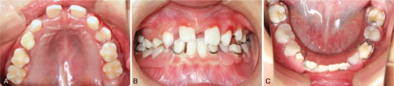
The extraoral examination presented no abnormity. Intraorally, the patient was in mixed dentition and the oral mucosa was normal. Examination revealed the presence of following teeth: #16, 55, 54, 53, 11, 21, 63, 64 65, 26, 36, 74, 31, 32, 41, 42, and 46 (Fig. 1). Tooth #24 and 43 were partially erupted. Her maxillary 2nd primary molars looked slightly abnormal in shape, with a comparatively larger cusp of Carabelli than usual. All other erupted teeth were regular in shape and color. All teeth were small in size with evident interdental spaces. The patient also had an anterior crossbite on her right maxillary canine. For exact diagnosis, we made a plaster cast and measured each erupted tooth 3 times including mesiodistal and labiolingual dimensions. The average value was obtained to improve measurement precision. All teeth were a smaller size than normal in every dimension (Tables 1 and 2). Moreover, the distal-occlusal caries of her mandible left 1st primary molar had periapical periodontitis, and the permanent successor had partially erupted. Furthermore, her mandible left 2nd primary molar had a deep caries lesion.
Figure 1.

Intraoral clinical photographs, including views of: (A) upper arch; (B) anterior; (C)lower arch.
Table 1.
Comparison of mesiodistal and buccolingual/labiolingual dimensions with an anatomic average value for the maxillary and mandibular permanent teeth (unit of measurement: millimeters).

Table 2.
Comparison of mesiodistal and buccolingual/labiolingual dimensions with an anatomic average value for the maxillary and mandibular primary teeth (unit of measurement: millimeters).

To verify whether this patient had tooth deficiency, a panoramic radiograph was taken (Fig. 2). It revealed that tooth #17, 13–15, 23–25, 27, 37, 33–35, 43–45, and 47 were unerupted or partially erupted. Moreover, the permanent teeth germs for the bilateral maxillary lateral incisor were congenitally absent. We tested the mesiodistal dimension of the erupted teeth from panoramic radiograph and casts. Thus, we obtained a magnified X-ray film and further predicted the mesiodistal dimensions of the unerupted teeth. Every measurement was also taken 3 times and the average value was obtained. The outcomes showed that all unerupted teeth were also smaller than usual (Table 1). Additionally, the panoramic radiograph revealed her maxillary 1st permanent molar had a single root and single canal bilaterally. However, the patient in our case did not present any endodontic symptoms. Cone-beam computerized tomography (CBCT) and periapical examinations were not conducted to avoid over-treatment.
Figure 2.

Dental panoramic radiograph of the patient.
Based on radiological and clinical measurements, the patient was diagnosed with simultaneous true generalized microdontia, hypodontia, and a rare variation of maxillary 1st molar with a single root and single canal. Also, the patient had premature loss of mandibular primary molars and canines. At the current stage, we made a removable appliance to hold space for the early loss of mandibular molars and canines in this patient (Fig. 3). Her mandible left 1st primary molar was extracted and the left 2nd primary molar was filled with complex resin materials. A multi-disciplinary therapy plan will be carefully formed including orthodontics, dental implants, and esthetic restoration for this patient in the future. The patient and her guardian have provided informed consent for publication of the case.
Figure 3.
Removable appliance to hold space for mandibular primary molars and canines:(A) upper arch; (B) anterior; (C)lower arch.
3. Discussion
Dental abnormality in size, number, shape, and structure can occur at any stage of tooth development, affecting a patient's quality of life. The complex pathogenesis can be divided into genetic and environmental factors.[7] During tooth development, dental epithelial cells form enamel, while ectomesenchymal stem cells differentiate and generate dentine, cementum, pulp, and the periodontal ligament. The interaction of dental epithelium and mesenchyme influences the number, position and morphogenesis of teeth. Although the mechanism remains unclear, many studies have shown that genetic mutations, epigenetics and environmental factors contribute to variations in teeth.[7]
Microdontia is a type of dental anomaly in which teeth are smaller in size than normal. This abnormality can occur in permanent teeth and primary teeth. According to epidemiological studies, the prevalence of microdontia ranges from 1.5 to 2% and occurs more frequently in females than males.[9] It has been reported that the most affected position is maxillary lateral incisors.[10] Microdontia often manifests as ‘Peg-shaped’ teeth, which are called ‘conical teeth’.[9] Single tooth microdontia is often reported, while true generalized microdontia is rare and is often found as a part of syndrome, such as pituitary dwarfism, Gorlin-Chaudhry-Moss syndrome, and Williams's syndrome, among others.[4] To the best of our knowledge, before this case, only 1 patient was reported with non-syndromic microdontia involving the whole dentition.[4] The patient in our case did not have any abnormal systemic diseases and all relevant diseases were ruled out. The etiology of microdontia remains poorly understood. Genetic factors are regarded as the main cause determining the size of teeth,[11,12] but isolated cases also can be found without any notable causes.[13] Treatment for microdontia usually needs esthetic restoration. Orthodontics may be necessary to close gaps between teeth.[5]
Hypodontia is common and affects permanent teeth more often, with prevalence ranging from 2.6 to 11.3% (excluding the 3rd molars).[14] Although it usually occurs alone, hypodontia can be found along with other dental abnormalities such as supernumerary, microdontia, short roots, enamel hypoplasia, and taurodontisim.[15] The simultaneous presence of hypodontia and single tooth microdontia has been frequently reported in some studies.[16] According to previous studies, the position of hypodontia varies according to race. In Europe, the maxillary 2nd premolar is the most reported position, while in America it is the maxillary lateral incisor.[17] The congenic absence of mandibular lateral incisors in Asians occurs more frequently. Although a larger proportion of hypodontia tends to occur unilaterally, the bilateral absence of lateral incisors is more common.[15]
It is suggested more than 300 genes regulate and control the process of tooth development.[18] The most well-studied genes related to hypodontia include AXIN2, MSX1, and PAX9.[19–21] These genes also participate in the development of other organs, thus hypodontia can occur with other syndromes such as cleft lip and palate, hereditary hypohidrotic ectodermal dysplasia and Down syndrome. Environment is another risk factor leading to hypodontia. It has been shown that maternal rubella virus infection, maxillofacial trauma, the use of thalidomide and chemotherapy drugs may lead to hypodontia.[15]
We report the simultaneous presence of true generalized microdontia, hypodontia, and a rare variation of the maxillary 1st molar with a single root and single canal in the same patient without any systemic diseases. The size of the teeth in our case was smaller than those reported in Bargale and Kiran's case. As reported, the absence of maxillary incisors bilaterally and the small size of all teeth severely affected the patient's masticatory functions and facial esthetics. Furthermore, the rare variation of root canal anatomy in our case requires further tracking. At present, the patient has complied well with instructions to wear a removable space maintainer, which will help prevent mesial migration of the permanent 1st molars. In the future, a multi-disciplinary therapy plan should be carefully made including orthodontics, dental implant and esthetic restoration.
4. Conclusion
Hypodontia is a common dental anomaly, while true generalized microdontia without any systemic diseases and variation of the maxillary 1st molar with a single root and single canal is quite unique. To the best of our knowledge, this is the 1st case reporting the simultaneous occurrence of these dental anomalies in the same patient. The etiology remains unclear and further studies are required to explore it. As for a treatment program, this patient requires cooperation among many subjects in the future.
Author contributions
Conceptualization: Yan Wang.
Investigation: Fangjie Zhou.
Supervision: Yan Wang.
Validation: Fangjie Zhou.
Visualization: Fangjie Zhou.
Writing – original draft: Yuan Chen, Luxian Chen.
Writing – review & editing: Fangjie Zhou, Yiran Peng, Yan Wang.
Yan Wang orcid: 0000-0002-7136-9353.
Footnotes
Abbreviation: CBCT = cone-beam computerized tomography.
YC and FZ contributed equally to this article and should be regarded as co-first authors.
The authors have no funding and conflicts of interest to disclose.
Informed written consent was obtained from the patient for publication of this case report and accompanying images.
References
- [1].Shafer WG, Hine MK, Levy BM. A textbook of oral pathology. 1983;Philadelphia: W.B Saunders Co, 26p. [Google Scholar]
- [2].Villa A, Albonico A, Villa F. Hypodontia and microdontia: clinical features of a rare syndrome. J Can Dent Assoc 2011;77:115. [PubMed] [Google Scholar]
- [3].Opinya GN, Kaimenyi JT, Meme JS. Oral findings in Fanconi's anemia. A case report. J Periodontol 1988;59:461–3. [DOI] [PubMed] [Google Scholar]
- [4].Bargale SD, Kiran SD. Non-syndromic occurrence of true generalized microdontia with mandibular mesiodens – a rare case. Head & Face Med 2011;7:19. [DOI] [PMC free article] [PubMed] [Google Scholar]
- [5].Laverty DP, Thomas MBM. The restorative management of microdontia. Brit Dent J 2016;221:160. [DOI] [PubMed] [Google Scholar]
- [6].Nunn JH, Carter NE, Gillgrass TJ, et al. The interdisciplinary management of hypodontia: background and role of paediatric dentistry. Br Dent J 2003;194:245–51. [DOI] [PubMed] [Google Scholar]
- [7].Ezoddini AF, Sheikhha MH, Ahmadi H. Prevalence of dental developmental anomalies: a radiographic study. Community Dent Hlth 2007;24:140. [PubMed] [Google Scholar]
- [8].Saxena A, Avantika S, Anuja L. A rare case of maxillary first molar with single root and single canal diagnosed using spiral computed tomographic scan. J Indian Soc Pedod Prev Dent 2014;32:242–5. [DOI] [PubMed] [Google Scholar]
- [9].Guttal KS, Naikmasur VG, Bhargava P. Frequency of developmental dental anomalies in the Indian population. Eur J Dent 2010;4:263–9. [PMC free article] [PubMed] [Google Scholar]
- [10].Fekonja A. Prevalence of dental developmental anomalies of permanent teeth in children and their influence on esthetics. J Esthet Restor Dent 2017;(1.): [DOI] [PubMed] [Google Scholar]
- [11].Steinberg AG, Warren JF, Warren LM. Hereditary generalized microdontia. J Dent Res 1961;40:58–62. [Google Scholar]
- [12].Santosh P, Nidhi Y, Prashant P. Non-syndromic occurrence of multiple dental and skeletal anomalies: a rare case report and brief literature review. J Clin Diagn Res 2014;8:28–30. [DOI] [PMC free article] [PubMed] [Google Scholar]
- [13].Khan S, Gill D, Bassi GS. Management of microdont maxillary lateral incisors. Dent Update 2014;41:867–74. [Google Scholar]
- [14].O’Dowling IB, Mcnamara TG. Congenital absence of permanent teeth among Irish school-children. J Ir Dent Assoc 1990;36:136–8. [PubMed] [Google Scholar]
- [15].De Coster P, Marks L, Martens L, et al. Dental agenesis: genetic and clinical perspectives. J Oral Pathol Med 2009;38:1–7. [DOI] [PubMed] [Google Scholar]
- [16].Singhal P, Singhal P, Gupta R. True generalized microdontia and hypodontia with spondyloepiphyseal dysplasia. Case Reports in Dent 2013;2013:1–4. [DOI] [PMC free article] [PubMed] [Google Scholar]
- [17].Altug-atac AA, Dilek E. Prevalence and distribution of dental anomalies in orthodontic patients. Orthodontics 2012;131:510–4. [DOI] [PubMed] [Google Scholar]
- [18].Ritwik P, Kimberly K, Patterson Diagnosis of tooth agenesis in childhood and risk for neoplasms in adulthood. Ochsner J 2018;18:345–50. [DOI] [PMC free article] [PubMed] [Google Scholar]
- [19].Ye XQ, Attaie A. Genetic basis of nonsyndromic and syndromic tooth agenesis. J Pediatr Genet 2016;5:198–208. [DOI] [PMC free article] [PubMed] [Google Scholar]
- [20].Yin W, Bian Z. The gene network underlying hypodontia. J Dent Res 2015;94:878–85. [DOI] [PubMed] [Google Scholar]
- [21].Juuri E, Balic A. The biology underlying abnormalities of tooth number in humans. J Dent Res 2017;96:002203451772015. [DOI] [PubMed] [Google Scholar]