Abstract
Circular RNAs (circRNAs) are a large class of endogenous noncoding RNAs that regulate gene expression and mainly function as microRNA sponges. This study aimed to explore the aberrant expression of circRNAs in colorectal cancer (CRC). Using a circRNA microarray, we identified 892 differentially expressed circRNAs between six pairs of CRC and adjacent paracancerous tissues. Among them, hsa_circ_0007142 was significantly upregulated. Further analysis in 50 CRC clinical samples revealed that hsa_circ_0007142 upregulation was associated with poor differentiation and lymphatic metastasis of CRC. Bioinformatic analysis and luciferase reporter assay showed that hsa_circ_0007142 targeted miR-103a-2-5p in CRC cells. Moreover, the silencing of hsa_circ_0007142 by siRNAs decreased the proliferation, migration, and invasion of HT-29 and HCT-116 cells. Taken together, these findings suggest that hsa_circ_0007142 is upregulated in CRC and targets miR-103a-2-5p to promote CRC.
1. Introduction
Colorectal cancer (CRC) has the third highest incidence among cancers and is the fourth leading cause of cancer-related mortality worldwide [1]. Although several treatment options have achieved remarkable advances, the survival of CRC patients remains unsatisfactory, mainly due to the lack of effective biomarkers for stratification in personalized treatment. With the advent of precision medicine and next generation sequencing, it is urgent to identify valuable biomarkers for the diagnosis, prognosis, and therapy of CRC.
Circular RNAs (circRNAs) are noncoding RNAs that play an important role in regulating gene expression and function [2]. Compared with regular linear RNAs, circRNAs are produced with covalently linked ends, leading to increased stability [3]. Accumulating data have shown that circRNAs are aberrantly expressed in many tumors [4–8]. CircRNAs mainly serve as microRNA (miRNA) sponges to regulate miRNAs, such as the interaction of CDR1as with miR-7 in cervical carcinoma [9], the sponge function of circRNA-100338 for miR-141-3p in hepatocellular carcinoma (HCC) [10], and circRNA_100269 as the sponge for miR-630 in gastric cancer (GC) [11]. In particular, dysregulated circRNAs play important role in CRC via the regulation of multiple miRNAs [12, 13]. Furthermore, circRNAs could serve as transcriptional enhancers or competitors with linear splicing to regulate host gene expression in multiple diseases [14–17]. Many circRNAs have been reported to be potential biomarkers of CRC [18]. However, the function of these dysregulated circRNAs in CRC remains to be further investigated. In our previous study, we performed a circRNA microarray analysis with a panel of six CRC tissues and six normal tissues [19]. In this study, we focused on circRNA hsa_circ_0007142 because it showed the second highest upregulation in CRC in previous study [19]. We first confirmed the upregulation of hsa_circ_0007142 in CRC tissues by RT-qPCR analysis and analyzed the association of hsa_circ_0007142 with clinicopathological aspects of CRC. Next we investigated the functional role of hsa_circ_0007142 in the proliferation, migration, and invasion of CRC cells.
2. Materials and Methods
2.1. Tissue Specimens and Clinical Data
The present study was approved by the Ethics Committee of Huai'an First People's Hospital, Nanjing Medical University (Huai'an, China), and written informed consent was obtained from each patient. The tissue samples of CRC and paired adjacent paracancerous tissues were obtained from CRC patients at Huai'an First People's Hospital. All patients did not receive prior radiotherapy and chemotherapy, and CRC was confirmed by experienced pathologists. After surgery, the tissues were quickly snap-frozen and stored at -80°C until further analysis.
2.2. Cell Culture
Human colorectal cancer HCT-116, HT-29, and LoVo cells and normal human enteral epithelial (HCO) cells were obtained from the Shanghai Institutes for Biological Sciences (Shanghai, China) and cultured in Roswell Park Memorial Institute (RPMI) 1640 medium (Hyclone, Logan, UT, USA) supplemented with 10% FBS, 100 units/mL of penicillin, and 100 mg/mL of streptomycin at 37°C in an incubator with 5% CO2.
2.3. Reverse Transcription Quantitative PCR (RT-qPCR)
Total RNA was isolated from CRC tissues and cells using TRIzol reagent as described previously [20]. For circRNA analysis, cDNAs were synthesized with 1 μg of total RNA using random primers. For miRNAs, specific primers were used for cDNA synthesis. The expression levels of circRNAs or mRNA were determined using RT-qPCR, with GAPDH as an internal control. For miRNA expression, U6 was used as an internal control for normalization. All experiments were independently conducted in triplicate, and the 2−ΔΔCT method was used to analyze the data. The classification of CRC patient grouping was derived from the value of 2−ΔΔCT and the cut-off of the hsa_circ_0007142-high group was set as 1. The sequences of the primers are listed in Table 1.
Table 1.
The sequences for primers and siRNAs.
| Primers used for qRT-PCR | ||
| GAPDH F | GGGAGCCAAAAGGGTCAT | |
| GAPDH R | GAGTCCTTCCACGATACCAA | |
| hsa_circ_0007142 F | GAACTCTGCCTCAGGATGAA | |
| hsa_circ_0007142 R | AACGTGTAACCTCGGTACCA | |
| U6 | F | CTCGCTTCGGCAGCACA |
| U6 | R | AACGCTTCACGAATTTGCGT |
| U6 | RT | GTCGTATCCAGTGCAGGGTCCGAGGTAT |
| TCGCACTGGATACGACCAAATATGGAAC | ||
| miR-103a-2-5p | F | GCGCGAGCTTCTTTACAGTGCT |
| miR-103a-2-5p | R | ATCCAGTGCAGGGTCCGAGG |
| miR-103a-2-5p | RT | GTCGTATCCAGTGCAGGGTCCGA |
| GGTATTCGCACTGGATACGACCA AGGC | ||
| siRNAs oligonucleotides | ||
| si-circ_0007142 | GGAAACAGCTTTTTATAAC | |
2.4. Cell Transfection
The control and si-hsa_circ_0007142 siRNAs (mixtures of three siRNAs targeting different sites of hsa_circ_0007142) were designed and synthesized by RiboBio (Guangzhou, China; Table 1). HT-29 and HCT116 cells were seeded in six-well plates at a density of 5×10⁵/well and cultured to a confluency of 50-60% and then were transfected with control or gene specific siRNAs using Lipofectamine 2000 (Invitrogen, Carlsbad, CA, USA).
2.5. Cell Proliferation Assay
The Cell Counting Kit-8 (CCK-8, Dojindo, Japan) assay was performed to evaluate CRC cell proliferation. Briefly, 48 h after transfection, cells were seeded in 96-well plates at a density of 5×10³/well, and cell viability was measured on 1, 2, 3, and 4 days. CCK-8 reagent was added into each well and maintained at 37°C for 2 h. The OD value at 450 nm was measured using a microplate reader (Bio-Rad, Hercules, CA, USA).
2.6. Colony Formation Assay
Transfected cells were seeded into 6-well plates and cultured for 15 days. Then the cells were fixed with methanol and stained with 0.5% crystal violet (Beyotime Biotechnology) for 30 min. Colonies with more than 10 cells were counted under a light microscope.
2.7. Cell Migration and Invasion Assay
A 24-well Transwell insert with 8 μm pore (Corning, NY, USA) was used for the cell migration and invasion assays. 48 h after transfection, 1×10⁵ cells were plated in the upper chamber and 500 μl of medium was added to the lower chamber for the migration assay. For the invasion assay, the upper layer of membrane inserts was added with 40 μg of Matrigel per well and incubated at 37°C for 1 h. After 48 h, cells that migrated to the lower compartment through the coated membrane were fixed with methanol, stained with 1% crystal violet, and quantified under a microscope. All assays were performed in triplicate.
2.8. Luciferase Assay
The plasmids carrying the fragment of either the wild-type or the mutant hsa_circ_0007142 sequence of the predicted miR-103a-2-5p binding sites were constructed by Geneseed (Guangzhou, China). HT-29 cells were cotransfected with the plasmids and miR-103a-2-5p mimic using Lipofectamine 2000 (Invitrogen, Carlsbad, CA, USA). After 48 h, cells were collected and luciferase activity was determined using the Dual Luciferase Reporter Assay System (Promega, Madison, WI, USA).
2.9. Statistical Analysis
All data were presented as mean ± standard deviation (SD) and analyzed using SPSS 20.0 software (IBM, USA). Differences between groups were analyzed using Student's t-test, Wilcoxon test, or X2-test. A two-tailed P-value <0.05 was considered statistically significant.
3. Results
3.1. hsa_circ_0007142 Is Upregulated and Correlates with the Clinicopathological Features of CRC Patients
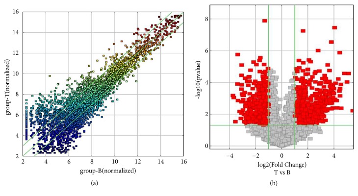
To assess the expression of circRNAs in CRC, the differential expression of circRNAs between six CRC tissues and six paracancerous tissues was examined by circRNA microarray. A total of 892 differentially expressed circRNAs were found in CRC, including 412 upregulated and 480 downregulated (fold >2, P<0.05; Figures 1(a) and 1(b)). The top 10 upregulated and downregulated circRNAs are listed in Table 2. The five potential target miRNAs of each of the top 10 upregulated and downregulated circRNAs were predicted by Arraystar software according to TargetScan and miRanda database (Table 3). Based on the expression level, the specificity of primers, the efficiency of siRNA-interference, and the function of potentially targeted miRNAs, we chose hsa_circ_0007142 derived from linear RNA DOCK1 for further investigation.
Figure 1.
Comparison of circRNA expression profiles between CRC and paired pericancerous tissues. (a) Scatter plots for assessing the difference in the expression of circRNAs among samples (T for CRC and B for adjacent tissues). The values of each group were plotted on the X and Y axes (log2 scaled). The middle green line represented no difference between two groups, while the flank green lines indicated 2-fold changes. The circRNAs beyond the lines referred to > 2-fold changes between two groups. (b) Volcano plots for differential circRNA expression between two groups. The vertical lines indicated 2 folds (log2 scaled), and the horizontal line represented P of 0.05 (log10 scaled). The red dots represented differentially expressed circRNAs with statistical significance.
Table 2.
The top ten upregulated and downregulated circRNAs in CRC ranked by fold changes in microarray data.
| CircRNA ID | CircRNA type | Chrom | Gene symbol | Fold change | Regulation | P value |
|---|---|---|---|---|---|---|
| hsa_circ_0000072 | exonic | chr1 | OMA1 | 43.4572991 | up | 0.005985797 |
| hsa_circ_0007142 | exonic | chr10 | DOCK1 | 34.9353568 | up | 2.71168E-05 |
| hsa_circ_0008812 | exonic | chr9 | RAD23B | 32.0813361 | up | 0.00009244 |
| hsa_circ_0050514 | exonic | chr19 | UBA2 | 27.7446201 | up | 2.68425E-05 |
| hsa_circ_0085923 | exonic | chr8 | PLEC | 23.5020249 | up | 0.004162563 |
| hsa_circ_0087855 | exonic | chr9 | RAD23B | 23.161069 | up | 0.000133271 |
| hsa_circ_0005954 | exonic | chr6 | AMD1 | 22.1403464 | up | 0.00069505 |
| hsa_circ_0005050 | exonic | chr2 | XPO1 | 22.0953662 | up | 0.000244329 |
| hsa_circ_0005281 | exonic | chr17 | TBCD | 21.7534364 | up | 1.32686E-06 |
| hsa_circ_0000994 | exonic | chr2 | SLC8A1 | 21.3375077 | up | 0.000346993 |
| hsa_circ_0072279 | exonic | chr5 | NUP1 | 13.3423298 | down | 0.000659178 |
| hsa_circ_0001704 | intragenic | chr7 | - | 12.7785132 | down | 0.000891495 |
| hsa_circ_0035626 | exonic | chr15 | RPS27L | 11.6798313 | down | 0.029341895 |
| hsa_circ_0001525 | intronic | chr5 | - | 10.49926 | down | 0.000123931 |
| hsa_circ_0005904 | exonic | chr2 | DCAF17 | 10.3297308 | down | 1.74038E-06 |
| hsa_circ_0008367 | exonic | chr9 | IARS | 10.183079 | down | 0.00136856 |
| hsa_circ_0047303 | exonic | chr18 | ZNF521 | 9.2977975 | down | 0.006717343 |
| hsa_circ_0087565 | exonic | chr9 | ZNF169 | 8.7255455 | down | 0.018358601 |
| hsa_circ_0000488 | intronic | chr13 | - | 8.4844294 | down | 0.000260793 |
| hsa_circ_0031569 | exonic | chr14 | HEATR5A | 8.432469 | down | 0.00854826 |
CircRNA ID: The circRNA ID can be found in circBase (http://www.circbase.org/). Gene symbol represents the liner RNA where
circRNAs generated from. “-” indicates that circRNA microarray did not provide the gene symbol of circRNA derived from intragenic or intronic types.
Fold Change: calculated from the ratio of the two groups (normal tissues versus CRC tissues). P-value: computed from paired t-test.
Table 3.
Five target miRNAs of differentially expressed circRNAs in microarray data.
| CircRNA ID | Regulation | MRE |
|---|---|---|
| hsa_circ_0000072 | up | hsa-miR-136-5p, hsa-miR-606, hsa-miR-145-5p, hsa-miR-153-5p, hsa-miR-302d-5p |
| hsa_circ_0007142 | up | hsa-miR-651-3p,hsa-miR-103a-2-5p,hsa-miR-744-3p,hsa-miR-96-3p,hsa-miR-128-3p |
| hsa_circ_0008812 | up | hsa-miR-138-5p,hsa-miR-325,hsa-miR-593-3p,hsa-miR-512-3p,hsa-miR-766-5p |
| hsa_circ_0050514 | up | hsa-miR-433-3p,hsa-miR-215-3p,hsa-miR-34b-5p,hsa-miR-23b-3p,hsa-miR-891a-5p |
| hsa_circ_0085923 | up | hsa-miR-580-5p,hsa-miR-198,hsa-miR-656-5p,hsa-miR-219a-5p,hsa-miR-758-5p |
| hsa_circ_0087855 | up | hsa-miR-138-5p,hsa-miR-325,hsa-miR-593-3p,hsa-miR-512-3p,hsa-miR-766-5p |
| hsa_circ_0005954 | up | hsa-miR-520f-3p,hsa-miR-618,hsa-miR-223-3p,hsa-miR-875-3p,hsa-miR-495-3p |
| hsa_circ_0005050 | up | hsa-miR-452-5p,hsa-miR-146b-5p,hsa-miR-146a-5p,hsa-miR-597-3p,hsa-miR-578 |
| hsa_circ_0005281 | up | hsa-miR-576-3p,hsa-miR-646,hsa-miR-219a-2-3p,hsa-miR-181b-2-3p,hsa-miR-181b-3p |
| hsa_circ_0000994 | up | hsa-miR-27b-3p,hsa-miR-27a-3p,hsa-miR-373-5p,hsa-miR-335-3p,hsa-miR-628-5p |
| hsa_circ_0072279 | down | hsa-miR-665,hsa-miR-874-5p,hsa-miR-188-3p,hsa-miR-29b-2-5p,hsa-miR-1271-3p |
| hsa_circ_0001704 | down | hsa-miR-20b-3p,hsa-miR-641,hsa-miR-766-3p,hsa-miR-661,hsa-miR-625-5p |
| hsa_circ_0035626 | down | hsa-miR-619-3p,hsa-miR-653-5p,hsa-miR-660-3p,hsa-miR-548d-5p,hsa-miR-200a-3p |
| hsa_circ_0001525 | down | hsa-let-7i-5p,hsa-let-7g-5p,hsa-miR-619-5p,hsa-let-7f-5p,hsa-miR-98-5p |
| hsa_circ_0005904 | down | hsa-let-7g-5p,hsa-let-7i-5p,hsa-miR-98-5p,hsa-miR-141-3p,hsa-miR-545-3p |
| hsa_circ_0008367 | down | hsa-miR-330-5p,hsa-miR-216a-3p,hsa-miR-342-5p,hsa-miR-891a-3p,hsa-miR-326 |
| hsa_circ_0047303 | down | hsa-miR-149-3p,hsa-miR-17-3p,hsa-miR-1301-3p,hsa-miR-509-5p,hsa-miR-516b-5p |
| hsa_circ_0087565 | down | hsa-miR-766-3p,hsa-miR-612,hsa-miR-338-3p,hsa-let-7g-3p,hsa-let-7a-2-3p |
| hsa_circ_0000488 | down | hsa-miR-519a-3p,hsa-miR-519b-3p,hsa-miR-28-5p,hsa-miR-130b-3p,hsa-miR-708-5p |
| hsa_circ_0031569 | down | hsa-miR-128-3p,hsa-miR-30c-2-3p,hsa-miR-216a-3p,hsa-miR-585-5p,hsa-miR-140-3p |
MRE: miRNA response element.
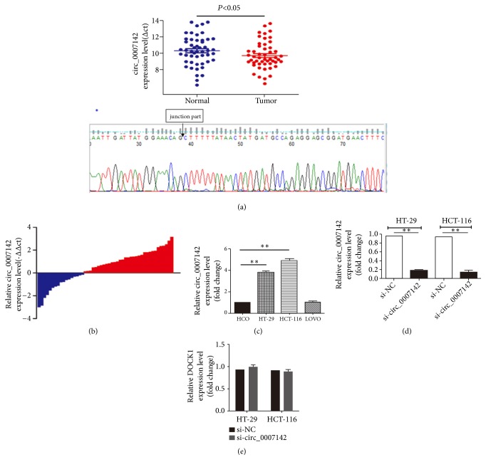
In order to validate the upregulation of hsa_circ_0007142 in CRC, we examined hsa_circ_0007142 expression level by RT-qPCR in 50 clinical CRC specimens and pericancerous tissues. Compared to adjacent noncancerous tissues, CRC tissues had a significantly higher hsa_circ_0007142 expression (P<0.05, Figures 2(a) and 2(b)). Sequencing results of PCR products confirmed the expression of hsa_circ_0007142 (Figure 2(a)). In addition, we analyzed the association of hsa_circ_0007142 expression with CRC clinical features and found that high expression of hsa_circ_0007142 was associated with the differentiation (P=0.008) and lymphatic metastasis of CRC (P=0.037) (Table 4). These results suggest the important role of hsa_circ_0007142 in the progression of CRC.
Figure 2.
hsa_circ_0007142 expression is elevated in CRC tissues and cells. (a) hsa_circ_0007142 expression in CRC tissues (n=50) and paired pericancerous tissues (n=50) was determined by PCR and normalized to GAPDH. Sequencing results confirmed PCR products, especially the junction part. (b) The tissues were divided into two groups. Positive –ΔΔcT indicated high hsa_circ_0007142 expression, while negative -ΔΔCT indicated low hsa_circ_0007142 expression. (c) Relative hsa_circ_0007142 expression in HT-29, HCT116, LoVo, and HCO cells. (d) Relative hsa_circ_0007142 expression level in HT-29 and HCT-116 cells transfected with circ_0007142 (si-circ_0007142) or control siRNA (si-NC). (e) Relative DOCK1 mRNA expression in HT-29 and HCT116 cells transfected with si-circ_0007142 or si-NC. Error bars represented mean ± standard deviation (SD). ∗P<0.05 and ∗∗P<0.01. ΔCT is the difference between the CT value of hsa_circ_0007142 and CT value of GAPDH. ΔΔCT is the difference between the ΔCT of CRC tissues and the ΔCT of adjacent normal tissues.
Table 4.
Correlation between hsa_circ_0007142 expression and clinicopathological characteristics of CRC.
| Clinical Parameter | Hsa_circ_0007142 (2−ΔΔCT) | Chi-squared test | |
|---|---|---|---|
| High group | Low group | P value | |
| (≥1, n=33) | (<1, n=17) | ||
| Gender | 0.623 | ||
| Male | 21 | 12 | |
| Female | 12 | 5 | |
| Age (years) | 0.209 | ||
| ≤50 | 6 | 7 | |
| 50–70 | 15 | 6 | |
| > 70 | 12 | 4 | |
| Tumor size | 0.577 | ||
| ≤3 cm | 8 | 5 | |
| 3–5 cm | 17 | 10 | |
| >5 cm | 8 | 2 | |
| Differentiation | 0.008∗∗ | ||
| Poor | 2 | 6 | |
| Moderate | 24 | 11 | |
| Well | 7 | 0 | |
| Lymphatic metastasis | 0.037∗ | ||
| Nx&N0 | 13 | 12 | |
| N1&N2 | 20 | 5 | |
| Tumor stage | 0.136 | ||
| I–II | 19 | 6 | |
| III–IV | 14 | 11 | |
| Distant metastasis | 0.11 | ||
| No | 33 | 15 | |
| Yes | 0 | 2 | |
∗P<0.05, ∗∗P<0.01. ΔCT is the difference between the CT value of hsa_circ_0007142 and of GAPDH.
ΔΔCT is the difference between the ΔCT of CRC tissues and the ΔCT of adjacent normal tissues.
3.2. hsa_circ_0007142 Promotes the Proliferation of CRC Cells
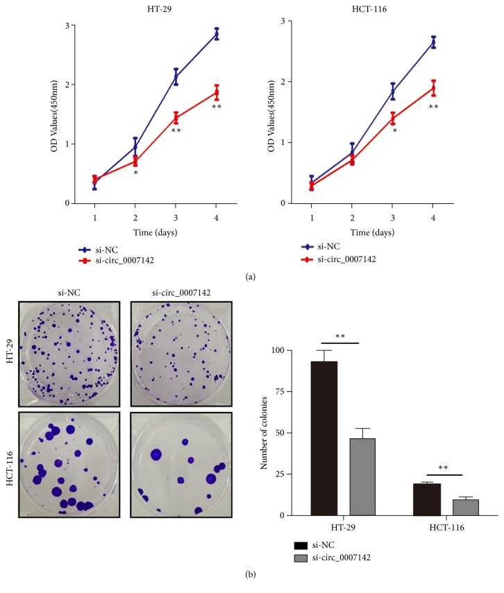
To explore the role of hsa_circ_0007142 in CRC, first we compared its expression level in CRC cell lines HCT-116, HT-29, and LoVo with normal enteral epithelial cell line HCO. The expression of hsa_circ_0007142 was significantly higher in HCT-116 and HT-29 cells than in HCO cells, but there was no significant difference between HCO and LoVo cells (Figure 2(c)). Next, loss-of-function assays were performed by transfecting specifically designed siRNAs or negative control siRNA into HT-29 and HCT-116 cells. We found that hsa_circ_0007142 siRNA significantly decreased hsa_circ_0007142 in both HCT-116 and HT-29 cell lines (Figure 2(d)). In order to exclude the possibility of siRNAs abating DOCK1, which produces hsa_circ_0007142 during the transcription, the expression level of DOCK1 mRNA was examined. As expected, no difference in DOCK1 expression was observed between HCT-116 and HT-29 cells transfected with hsa_circ_0007142 and negative control siRNAs (Figure 2(e)). CCK-8 assay showed that hsa_circ_0007142 knockdown by siRNA significantly inhibited the proliferation of HCT-116 and HT-29 cells in a time dependent manner (Figure 3(a)). In addition, colony formation assay showed that the colony forming ability of HCT-116 and HT-29 cells transfected with hsa_circ_0007142 siRNA was significantly reduced compared to cells treated with control siRNA (Figure 3(b)). Collectively, these results suggest that hsa_circ_0007142 promotes the proliferation of CRC cells.
Figure 3.
hsa_circ_0007142 knockdown inhibited CRC cell proliferation. (a) CCK8 assay of the viability of HT-29 and HCT116 cells transfected with circ_0007142 (si-circ_0007142) or control siRNA (si-NC). (b) Colony-forming assay of HT-29 and HCT-116 cells transfected with si-circ_0007142 or si-NC. Data were presented as the mean ± SD of three independent experiments. ∗P<0.05, ∗∗P<0.01.
3.3. hsa_circ_0007142 Enhances the Migration and Invasion of CRC Cells
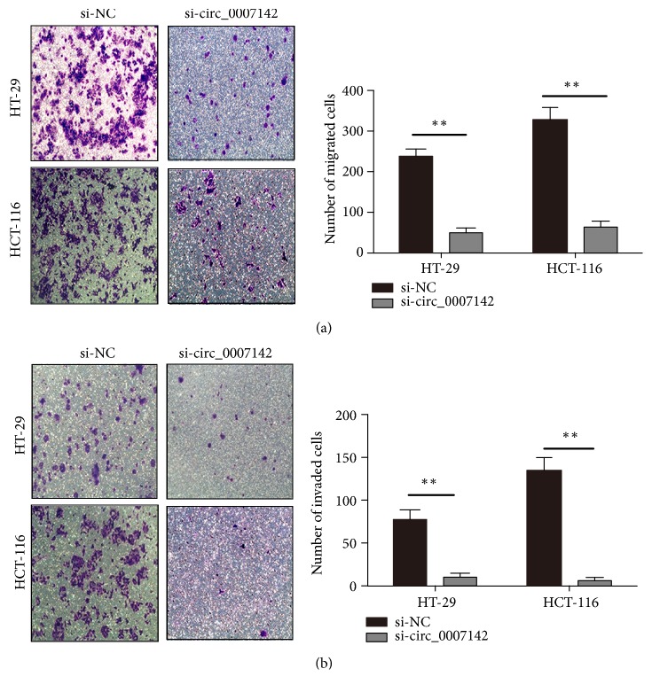
Since hsa_circ_0007142 upregulation was associated with lymphatic metastasis of CRC, next we investigated the role of hsa_circ_0007142 in the migration and invasion of CRC cells. Chamber assay showed that, compared to control siRNA groups, the knockdown of hsa_circ_0007142 by siRNA effectively decreased the invasion (Figure 4(a)) and migration (Figure 4(b)) of both HCT-116 and HT-29 cells.
Figure 4.
hsa_circ_0007142 knockdown inhibited CRC cell migration and invasion. Transwell assays on the migration (a) and invasion (b) of HT-29 and HCT-116 cells transfected with si-circ_0007142 or si-NC. ∗∗P<0.01; magnification ×100.
3.4. MiR-103a-2-5p Is the Direct Target of hsa_circ_0007142
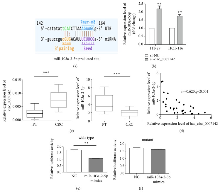
Arraystar software revealed that hsa_circ_0007142 might target miR-103a-2-5p (Figure 5(a)). As expected, significant increase in miR-103a-2-5p expression was found in cells transfected with hsa_circ_0007142 siRNA (Figure 5(b)). Next, the expression of miR-103a-2-5p and hsa_circ_0007142 in 30 pairs of CRC and adjacent noncancerous tissues was detected by RT-qPCR. We found that the expression of hsa_circ_0007142 was significantly upregulated while the expression of miR-103a-2-5p was significantly downregulated in CRC tissues compared to paired pericancerous tissues (PT) (Figure 5(c)). Moreover, the expression of miR-103a-2-5p was negatively correlated with hsa_circ_0007142 expression (P<0.01, r = -0.623, Figure 5(d)). Finally, luciferase reporter assay showed that luciferase activity significantly decreased in HT-29 cells cotransfected with the plasmids of wild-type hsa_circ_0007142 and miR-103a-2-5p mimics, compared to cells cotransfected with plasmids of mutant hsa_circ_0007142 and miR-103a-2-5p mimics (Figure 5(e)). In contrast, there was no significant difference in luciferase activity in HT-29 cells cotransfected with miR-103a-2-5p mimics and plasmids of mutant hsa_circ_0007142, where the miR-103a-2-5p binding site was mutated (Figure 5(f)). Taken together, these results demonstrate that hsa_circ_0007142 targets miR-103a-2-5p in CRC cells.
Figure 5.
MiR-103a-2-5p is a target of hsa_circ_0007142. (a) The binding site of miR-103a-2-5p for hsa_circ_0007142 was predicted by circRNA microarray. (b) miR-103a-2-5p expression was determined by RT-qPCR in HT-29 and HCT-116 cells transfected with si-circ_0007142 or si-NC. (c) PCR analysis of hsa_circ_0007142 and miR-103a-2-5p expression in CRC tissues (n=30) and paired pericancerous tissues (PT, n=30). (d) The correlation of the expression of miR-103a-2-5p and hsa_circ_0007142 in CRC tissues (n=30) and paired pericancerous tissues (n=30). (e) The luciferase activity in HT-29 cells cotransfected with the plasmids of wild-type hsa_circ_0007142 and miR-103a-2-5p mimics. (f) The luciferase activity in HT-29 cells cotransfected with plasmids of mutant hsa_circ_0007142 and miR-103a-2-5p mimics. ∗P<0.05, ∗∗P<0.01.
4. Discussion
As endogenous noncoding RNAs, circRNAs exhibit a remarkable organization- and disease-specific characteristic, suggesting that circRNAs may serve as specific biomarkers for disease diagnosis and therapy [21]. A negative relationship between the universal reduction of circRNAs and CRC cell proliferation was observed [21]. Furthermore, circRNA ITCH inhibited esophageal squamous cell carcinoma via regulating Wnt/β-catenin pathway [22]. Moreover, circRNAs are involved in epithelial-mesenchymal transition [23]. Numerous studies have revealed that circRNAs play a crucial role in the initiation and progression of cancers through multiple mechanisms [24]. The well-studied function of circRNAs is acting as miRNA sponge to insulate miRNAs and terminate the regulation of their target genes [9, 25]. For example, circHIPK3, as a sponge of miR-124, could upregulate miR-124 target AQP3 expression in hepatocellular carcinoma [26]. Furthermore, circDOCK1 has been reported to inhibit the function of miR-196a-5p by binding miR-196a-5p and enhancing the expression of BIRC3 in OSCC [27]. With the rapid advance of deep RNA sequencing, an increasing number of aberrantly expressed circRNAs have been identified. However, the function of circRNAs in the initiation, progression, and maintenance of different cancers is far from being understood.
In present study, we performed circRNA microarray analysis and identified 892 differentially expressed circRNAs in CRC tissues versus paracancerous tissues. We focused on a distinctly upregulated circRNA hsa_circ_0007142, which was confirmed to be upregulated in CRC tissues and cells. The silencing of hsa_circ_0007142 by siRNAs decreased the proliferation, migration, and invasion of CRC cells. Importantly, hsa_circ_0007142 upregulation was correlated to the differentiation and lymphatic metastasis of CRC. These findings suggest that hsa_circ_0007142 might play an oncogenic role in CRC.
Furthermore, bioinformatics predicted that miR-103a-2-5p was a target of hsa_circ_0007142. While several studies suggested oncogenic role of miR-103a in CRC [28, 29], it was reported that miR-103a could inhibit gastric cancer cell proliferation, migration, and invasion [30]. In addition, miR-103a-2-5p had the capability to inhibit cell survival and colony formation by targeting PARP-1 [31]. Nevertheless, the role of miR-103a-2-5p in CRC remains unknown up to date. In this study we employed luciferase assay to confirm that miR-103a-2-5p was a target of hsa_circ_0007142. It should be pointed out that circRNAs usually target several miRNAs. For example, circRNA ITCH acted as the sponge of miR-7, miR-17, and miR-214 to inhibit esophageal squamous cell carcinoma [22]. Further studies are needed to identify other potential targets of hsa_circ_0007142 and better understand the role of hsa_circ_0007142 in CRC.
In summary, the expression of hsa_circ_0007142 is upregulated in CRC tissues and is associated with the differentiation and lymphatic metastasis of CRC. Furthermore, hsa_circ_0007142 promotes the proliferation and invasion of CRC probably by functioning as a sponge for miR-103a-2-5p. These findings may provide new clue for the strategies in the diagnosis and therapy of CRC.
Acknowledgments
This work was supported by grants from the Project Fund of Health Bureau of Jiangsu Province (H201358) and the Project Fund of Sci & Tech Bureau of Huai'an City (HAS2015001, HAS2012003, and HAP201206).
Data Availability
All data are available upon request.
Conflicts of Interest
The authors declare no conflicts of interest.
Authors' Contributions
Chang-li Zhu, Xiaofeng Sha, and Yuan Wang are co-first authors.
References
- 1.Siegel R. L., Miller K. D., Jemal A. Cancer statistics, 2017. CA: A Cancer Journal for Clinicians. 2017;67(1):7–30. doi: 10.3322/caac.21387. [DOI] [PubMed] [Google Scholar]
- 2.Chen L., Huang C., Wang X., Shan G. Circular RNAs in eukaryotic cells. Current Genomics. 2015;16(5):312–318. doi: 10.2174/1389202916666150707161554. [DOI] [PMC free article] [PubMed] [Google Scholar]
- 3.Chen L.-L., Yang L. Regulation of circRNA biogenesis. RNA Biology. 2015;12(4):381–388. doi: 10.1080/15476286.2015.1020271. [DOI] [PMC free article] [PubMed] [Google Scholar]
- 4.Qu S., Song W., Yang X., et al. Microarray expression profile of circular RNAs in human pancreatic ductal adenocarcinoma. Genomics Data. 2015;5:385–387. doi: 10.1016/j.gdata.2015.07.017. [DOI] [PMC free article] [PubMed] [Google Scholar]
- 5.Wang X., Zhang Y., Huang L., et al. Decreased expression of hsa_circ_001988 in colorectal cancer and its clinical significances. International Journal of Clinical and Experimental Pathology. 2015;8(12):16020–16025. [PMC free article] [PubMed] [Google Scholar]
- 6.Qin M., Liu G., Huo X., et al. Hsa_circ_0001649: a circular RNA and potential novel biomarker for hepatocellular carcinoma. Cancer Biomarkers. 2016;16(1):161–169. doi: 10.3233/cbm-150552. [DOI] [PubMed] [Google Scholar]
- 7.Liu M., Zhang X. An integrated analysis of mRNA-miRNA transcriptome data revealed hub regulatory networks in three genitourinary cancers. Biocell. 2017;41(1):19–26. [Google Scholar]
- 8.Song X., Zhang N., Han P., et al. Circular RNA profile in gliomas revealed by identification tool UROBORUS. Nucleic Acids Research. 2016;44(9, article no. e87) doi: 10.1093/nar/gkw075. [DOI] [PMC free article] [PubMed] [Google Scholar]
- 9.Hansen T. B., Jensen T. I., Clausen B. H., et al. Natural RNA circles function as efficient microRNA sponges. Nature. 2013;495(7441):384–388. doi: 10.1038/nature11993. [DOI] [PubMed] [Google Scholar]
- 10.Huang X., Huang Z., Xu Y., et al. Comprehensive circular RNA profiling reveals the regulatory role of the circRNA-100338/miR-141-3p pathway in hepatitis B-related hepatocellular carcinoma. Scientific Reports. 2017;7(1, article no. 5428) doi: 10.1038/s41598-017-05432-8. [DOI] [PMC free article] [PubMed] [Google Scholar]
- 11.Zhang Y., Liu H., Li W., et al. CircRNA_100269 is downregulated in gastric cancer and suppresses tumor cell growth by targeting miR-630. AGING. 2017;9(6):1585–1594. doi: 10.18632/aging.101254. [DOI] [PMC free article] [PubMed] [Google Scholar]
- 12.Hsiao K.-Y., Lin Y.-C., Gupta S. K., et al. Noncoding effects of circular RNA CCDC66 promote colon cancer growth and metastasis. Cancer Research. 2017;77(9):2339–2350. doi: 10.1158/0008-5472.CAN-16-1883. [DOI] [PMC free article] [PubMed] [Google Scholar]
- 13.Weng W., Wei Q., Toden S., et al. Circular RNA ciRS-7 — A promising prognostic biomarker and a potential therapeutic target in colorectal cancer. Clinical Cancer Research. 2017;23(14):3918–3928. doi: 10.1158/1078-0432.CCR-16-2541. [DOI] [PMC free article] [PubMed] [Google Scholar]
- 14.Yang W., Du W. W., Li X., Yee A. J., Yang B. B. Foxo3 activity promoted by non-coding effects of circular RNA and Foxo3 pseudogene in the inhibition of tumor growth and angiogenesis. Oncogene. 2016;35(30):3919–3931. doi: 10.1038/onc.2015.460. [DOI] [PubMed] [Google Scholar]
- 15.Kristensen L. S., Hansen T. B., Venø M. T., Kjems J. Circular RNAs in cancer: Opportunities and challenges in the field. Oncogene. 2018;37(5):555–565. doi: 10.1038/onc.2017.361. [DOI] [PMC free article] [PubMed] [Google Scholar]
- 16.Li J., Yang J., Zhou P., et al. Circular RNAs in cancer: novel insights into origins, properties, functions and implications. American Journal of Cancer Research. 2015;5(2):472–480. [PMC free article] [PubMed] [Google Scholar]
- 17.Qu S., Yang X., Li X., et al. Circular RNA: a new star of noncoding RNAs. Cancer Letters. 2015;365(2):141–148. doi: 10.1016/j.canlet.2015.06.003. [DOI] [PubMed] [Google Scholar]
- 18.Zhuo F., Lin H., Chen Z., Huang Z., Hu J. The expression profile and clinical significance of circRNA0003906 in colorectal cancer. OncoTargets and Therapy. 2017;10:5187–5193. doi: 10.2147/OTT.S147378. [DOI] [PMC free article] [PubMed] [Google Scholar]
- 19.Guo J.-N., Li J., Zhu C.-L., et al. Comprehensive profile of differentially expressed circular RNAs reveals that hsa_circ_0000069 is upregulated and promotes cell proliferation, migration, and invasion in colorectal cancer. OncoTargets and Therapy. 2016;9:7451–7458. doi: 10.2147/OTT.S123220. [DOI] [PMC free article] [PubMed] [Google Scholar]
- 20.Zhong J., Deng L., Jiang Y., Zou L., Yuan H., Tan S. Gene expression profiling of HepG2 cells after treatment with black tea polyphenols. Biocell. 2018;42(3):99–104. doi: 10.32604/biocell.2018.04915. [DOI] [Google Scholar]
- 21.Li P., Chen S., Chen H., et al. Using circular RNA as a novel type of biomarker in the screening of gastric cancer. Clinica Chimica Acta. 2015;444:132–136. doi: 10.1016/j.cca.2015.02.018. [DOI] [PubMed] [Google Scholar]
- 22.Li F., Zhang L., Li W., et al. Circular RNA ITCH has inhibitory effect on ESCC by suppressing the Wnt/β-catenin pathway. Oncotarget . 2015;6(8):6001–6013. doi: 10.18632/oncotarget.3469. [DOI] [PMC free article] [PubMed] [Google Scholar]
- 23.Conn S. J., Pillman K. A., Toubia J., et al. The RNA binding protein quaking regulates formation of circRNAs. Cell. 2015;160(6):1125–1134. doi: 10.1016/j.cell.2015.02.014. [DOI] [PubMed] [Google Scholar]
- 24.Meng S., Zhou H., Feng Z., et al. CircRNA: Functions and properties of a novel potential biomarker for cancer. Molecular Cancer. 2017;16(1, article no. 94) doi: 10.1186/s12943-017-0663-2. [DOI] [PMC free article] [PubMed] [Google Scholar]
- 25.Sumazin P., Yang X., Chiu H.-S., et al. An extensive MicroRNA-mediated network of RNA-RNA interactions regulates established oncogenic pathways in glioblastoma. Cell. 2011;147(2):370–381. doi: 10.1016/j.cell.2011.09.041. [DOI] [PMC free article] [PubMed] [Google Scholar]
- 26.Chen G., Shi Y., Liu M., Sun J. CircHIPK3 regulates cell proliferation and migration by sponging miR-124 and regulating AQP3 expression in hepatocellular carcinoma. Cell Death & Disease. 2018;9(2):p. 175. doi: 10.1038/s41419-017-0204-3. [DOI] [PMC free article] [PubMed] [Google Scholar]
- 27.Wang L., Wei Y., Yan Y., et al. CircDOCK1 suppresses cell apoptosis via inhibition of miR-196a-5p by targeting BIRC3 in OSCC. Oncology Reports. 2018;39(3):951–966. doi: 10.3892/or.2017.6174. [DOI] [PMC free article] [PubMed] [Google Scholar]
- 28.Caritg O., Navarro A., Moreno I., et al. Identifying high-risk stage ii colon cancer patients: a three-MicroRNA-based score as a prognostic biomarker. Clinical Colorectal Cancer. 2016;15(4):e175–e182. doi: 10.1016/j.clcc.2016.04.008. [DOI] [PubMed] [Google Scholar]
- 29.Fasihi A., M. Soltani B., Atashi A., Nasiri S. Introduction of hsa-miR-103a and hsa-miR-1827 and hsa-miR-137 as new regulators of Wnt signaling pathway and their relation to colorectal carcinoma. Journal of Cellular Biochemistry. 2018;119(7):5104–5117. doi: 10.1002/jcb.26357. [DOI] [PubMed] [Google Scholar]
- 30.Liang J., Liu X., Xue H., Qiu B., Wei B., Sun K. MicroRNA-103a inhibits gastric cancer cell proliferation, migration and invasion by targeting c-Myb. Cell Proliferation. 2015;48(1):78–85. doi: 10.1111/cpr.12159. [DOI] [PMC free article] [PubMed] [Google Scholar]
- 31.Dluzen D. F., Kim Y., Bastian P., et al. MicroRNAs modulate oxidative stress in hypertension through PARP-1 regulation. Oxidative Medicine and Cellular Longevity. 2017;2017:12. doi: 10.1155/2017/3984280.3984280 [DOI] [PMC free article] [PubMed] [Google Scholar]
Associated Data
This section collects any data citations, data availability statements, or supplementary materials included in this article.
Data Availability Statement
All data are available upon request.


