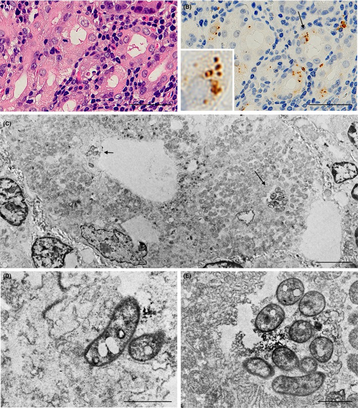
Figure 5.

Localization of Helicobacter pylori cells in secretory canaliculi of the infected parietal cells. Serial histologic sections of a sample with prominent H. pylori infection in parietal cells were used for (A) hematoxylin & eosin staining; (B) IHC with TMDU‐mAb (anti‐H. pylori); followed by (C) immuno‐electron microscopy. In b an arrow indicates H. pylori colonization and an inset shows higher magnification of the bacteria indicated by the arrow. (C) H. pylori colonization was observed in the secretory canaliculi of the parietal cells and is located near the entry (a short arrow) and in a deeper area (long arrow). (D and E) respectively show higher magnification of the H. pylori cells indicated in c by a short or long arrow. Note the intact and not denatured bacterial cells with a dense rim‐staining pattern corresponding to the distribution of the bacterial cell membrane‐bound lipopolysaccharide detected by the antibody. Bars: 50 µm (A and B), 5.0 µm (C) and 1.0 µm (D and E)
