. 2019 Jun 18;8:e47644. doi: 10.7554/eLife.47644

Figure 3. Structural features of the H. pylori Cag T4SS OMCC I-Layer and O-layer.

(A) Central axial slice view of the OMCC highlighting the position of the outer-layer (O-layer) and inner-layer (I-layer) colored in blue and red, respectively. Left panel, Surface representation; Right panel, Ribbon representation of secondary structure model. (B) Central axial slice view (left) and two cross sections of the OMCC highlighting the position of the spokes in O-layer (middle panel, blue, looking in from the outer membrane) and I-layer (right panel, red, looking out from the inner membrane).

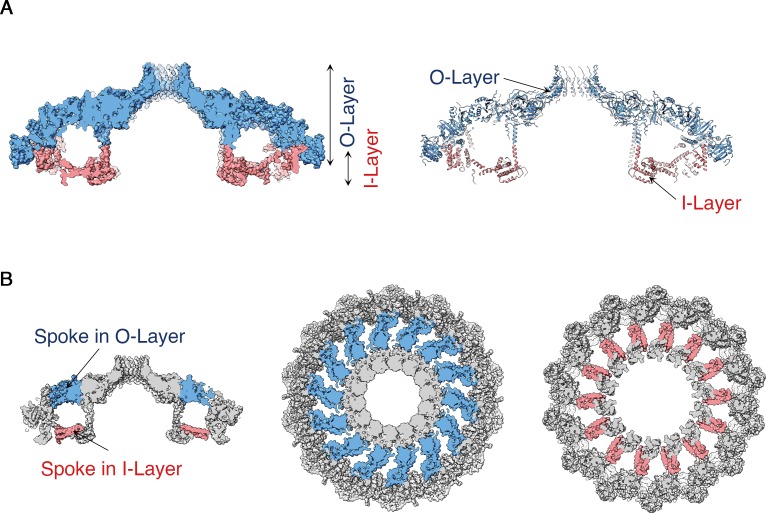
Figure 3.

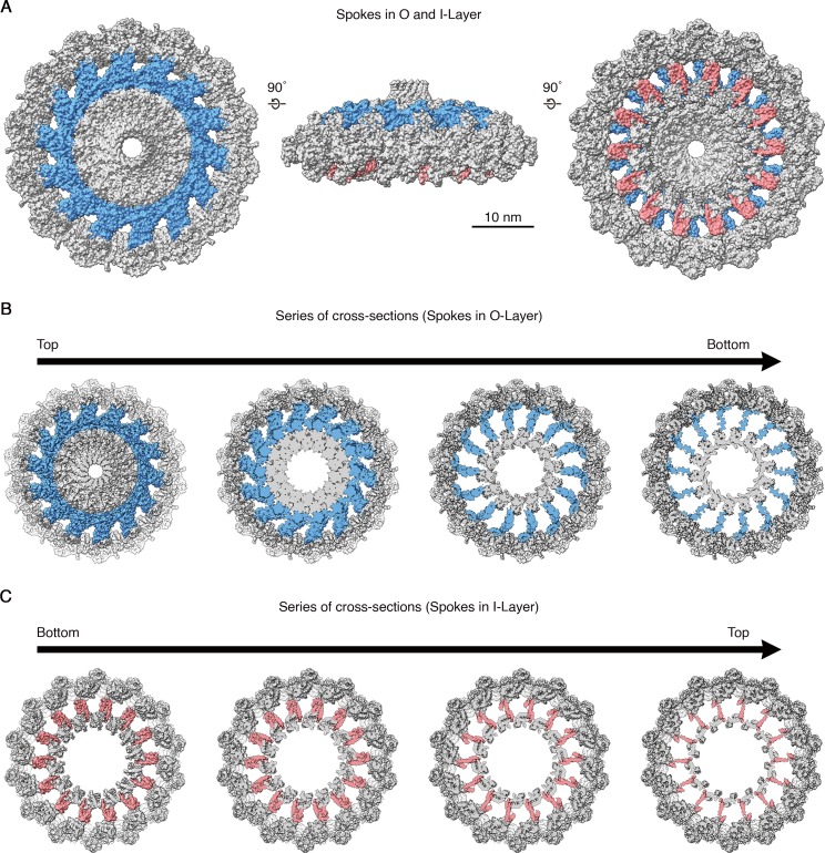
Figure 3—figure supplement 1. Spokes in O-layer and I-layer of OMCC.

Figure 3—figure supplement 1.

(A) Top (left), side (middle), and bottom (right) view of the OMCC highlighting the position of spokes in O-layer (blue) and I-layer (red). (B–C) Cross-section series of OMCC in different planes highlighting the position of spokes in O-layer. (B) View from the O-layer to I-layer (i.e. view looking in from the outer membrane towards the inner membrane) and C) View from the I-layer to O-layer (i.e. view looking out from inner membrane towards the outer membrane).