

Abstract
A 58-year-old male patient, with end-stage renal disease secondary to hypertension, underwent living-related kidney transplant at our transplant unit. The transplant surgery went uneventful and brisk urine output was recorded. Four hours after the transplant, the output suddenly dropped despite normal central venous pressure. Doppler scan revealed an extensive peri-allograft hematoma and high renal arterial resistive indices (RI). The patient was taken to the operating room where capsulotomy of the subcapsular hematoma was done. Postoperatively, the urine output restored to normal and the patient was sent home on the 5th post-operative day with adequately functioning renal graft. Surgical capsulotomy seems to be a valid approach in the management of such cases.
Keywords: Subcapsular hematoma, Doppler ultrasonography, Kidney transplantation, Renal graft hematoma
Introduction
Renal transplant is the established treatment of choice for patients with end-stage renal disease and provides better outcomes in terms of long-term survival and quality of life as compared to hemodialysis and peritoneal dialysis [1]. A rare complication in patients who undergo renal transplant is the development of subcapsular hematoma (SH) in the transplanted kidney. Subcapsular hematoma may result in oliguria, renal ischemia, Page kidney and even graft loss [2]. Ultrasound Doppler is a cheap, non-invasive and non-nephrotoxic way of diagnosing and monitoring the graft complications. It can safely be used within the first 24–48 h post-transplantation to detect the presence of and to monitor the evolution of subcapsular hematomas [3]. Different approaches used for the treatment of subcapsular hematoma include conservative management or surgical intervention including percutaneous drainage, capsulotomy or even nephrectomy [4, 5]. Here we have reported the case of a 58-year-old male patient who developed SH post-operatively with resultant oliguria for which surgical intervention was done. To the best of our knowledge, this is the first case of subcapsular hematoma post-renal transplant being reported in Pakistan.
Case report
A 58-year-old male patient, a known smoker with a BMI of 22.1 kg/m2, presented to us with end-stage renal disease secondary to hypertension. The patient had been on biweekly hemodialysis for 2 months pre-transplant. The patient had previous history of antiplatelet use. He had platelet count of 202 × 109/L and white cell count of 5.6 × 10⁹/L. His hemoglobin level was 9 g/dL, while hematocrit was 25.7%. His serum creatinine level was 6.81 mg/dL. Rest of the laboratory reports are mentioned in Table 1. Donor and recipient tissue typing revealed 5/10 antigen match and CDC cross match was negative. Flow cytometry was negative and Luminax Screen Test for HLA antibodies showed low level of IgG antibodies against class I and class II antigens. The immunosuppressive drugs used at induction were intravenous solumedrol 500 mg, oral valganciclovir yclovir 450 mg, oral mycophenolate mofetil 500 mg and antithymocyte globulin (ATG) 6 mg/kg. He received living-related kidney transplant from his son (aged 28 years).
Table 1.
Pertinent laboratory values
| TLC | 5.6 × 10⁹/L |
| Hemoglobin | 9 g/dL |
| Hematocrit | 25.7% |
| Platelets | 202 × 109/La |
| Creatinine | 6.81 mg/dLb |
| Sodium | 138 mEq/L |
| Potassium | 4.3 mEq/L |
| Bicarbonate | 22 mEq/L |
| Urea | 237 mg/dL |
| BUN | 111 mg/dL |
| Calcium | 8.7 mg/dL |
| Phosphorous | 6.4 mg/dL |
| PSA | 1.214 ng/mL |
| HBa1C | 4.9% |
| PT | 10.2 s |
| APTT | 21 s |
| INR | 1.0 |
aLitre
bDecilitre
Hand-assisted laparoscopic (HAL) donor nephrectomy was performed which went uneventful. The warm ischemia time was 2 min and 15 s whereas cold ischemia time was about 2 h. Recipient’s common iliac and external iliac arteries had calcified walls and after arteriotomy was done, a clot was visualized in the artery for which 5000 IU intravenous heparin was given intra-operatively. Rest of the transplant went uneventful and brisk urine output was recorded. The patient had a good urine output of 500–700 mL/h.
4 h after the transplant, the output suddenly dropped to 40 mL/h despite normal central venous pressure. His blood pressure and heart rate were normal and the patient was afebrile. Patient’s catheter was checked and was found to be alright. Normal saline bolus did not improve the condition.
Another attempt with 100 mg of intravenous Lasix along with a bolus of 200 mL of normal saline failed to improve urine output. Ultrasound Doppler was done after 1 h which showed an 83-mL subcapsular hematoma compressing the renal parenchyma with high renal arterial resistive indices (RI) (see Figs. 1, 2).
Fig. 1.
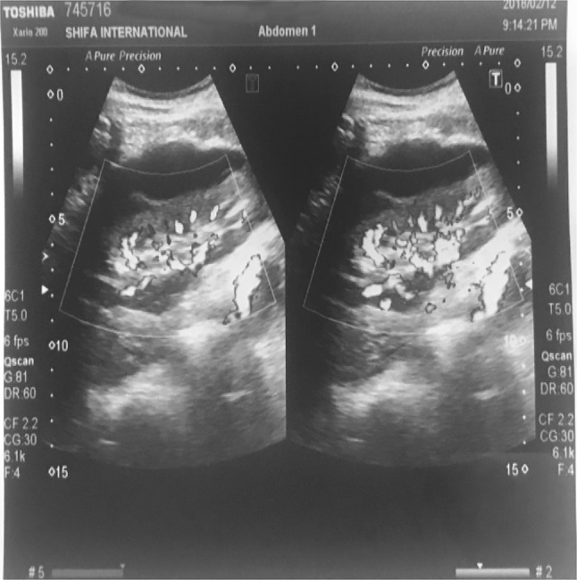
Subcapsular hematoma compressing the renal parenchyma
Fig. 2.
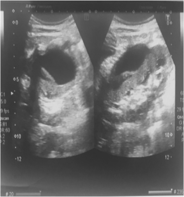
Subcapsular hematoma compressing the renal parenchyma
The patient was immediately shifted to the operating room and an emergency graft exploration was performed which showed a large subcapsular hematoma. Capsulotomy of the subcapsular hematoma was done. Multiple capsulotomy incisions were made. Serous fluid was released with pressure followed by clots and some amount of fresh blood. Urine output was restored to normal immediately after capsulotomy was done. The color of the kidney normalized and it became turgid immediately afterwards. Later on, the patient remained well and was sent home on the fifth post-operative day having serum creatinine of 0.76 mg/dL.
Discussion
Subcapsular renal hematoma pertains to hemorrhage from within the kidney that is contained by its fibrous capsule. This phenomenon was first described in an experimental model in the 1930 by Cromie and associates [9]. Acute hematomas may compress the renal parenchyma leading to delay in the graft function or even graft ischemia. Subcapsular hematomas may even result in hypertension induced by renal parenchymal ischemia (Page kidney) [6].
The most common cause of subcapsular hematoma is trauma. Trauma may occur during allograft procurement, peroperatively or during post-transplant biopsy. Tumors are another major cause and almost 50% of these tumors are malignant, with renal cell carcinoma being the most common of them [7]. Subcapsular hematoma may also result from lymphangiomas, periarteritis, anticoagulation therapy, and interventions such as renal biopsy and extracorporeal shockwave lithotripsy (ESWL) [8].
The signs and symptoms of subcapsular hematoma in renal allograft vary depending on the degree and duration of the bleeding. The clinical presentation of patients with a single kidney and renal allografts includes flank pain/tenderness, decreased urine output (as happened with our patient) or acute renal failure. A physical examination of the patient revealed diffuse moderate abdominal tenderness. Complete blood count and renal function tests were performed immediately which turned out to be within the normal limits. Doppler scan revealed an 83-mL extensive peri-allograft hematoma and high renal arterial resistive indices (RI), with an empty bladder and no dilation of the upper urinary tract.
Doppler scan can swiftly provide detailed information about the kidney sizes and echogenicity, the associated structures and any post-operative collections. Diagnosis of subcapsular hematoma is, however, best achieved with computerized tomography (CT) scan [9]. Angiography is another excellent imaging modality that can help identify the vascular abnormalities associated with subcapsular hematoma but is invasive, expensive and nephrotoxic. Magnetic resonance imaging (MRI) is another helpful diagnostic tool. Hematomas show high signal intensity with both T1-weighted and T2-weighted pulse sequences on an MRI. On scintigraphy, subcapsular hematomas appear as a “cold defect” [9]. In our patient, the sonographic findings provided quick and adequate information about the extent of subcapsular hematoma, so a CT scan was not performed.
The conservative treatment of subcapsular hematoma requires a strict follow-up with Doppler ultrasonography until the spontaneous resolution of the SH and recovery of graft function [9]. There have been other case reports of conservative line of therapy to be life threatening. Nurettin Ay et al. reported such a case of development of intra-operative subcapsular hematoma in renal allograft, which did not enlarge intra-operatively, for which close post-operative ultrasonography and hemodynamic follow-up were planned. The hematoma ruptured post-operatively and emergency re-exploration with subsequent extended capsulotomy had to be done [10].
In our patient, early diagnosis and timely capsulotomy helped swift recovery of the graft function.
Conclusion
We stress that in early diagnosed post-operative subcapsular renal graft hematomas, where there is decreased urine output, prompt surgical intervention can help reduce the risk of delayed complications such as impaired renal function and graft loss.
Acknowledgements
We thank Dr. Ayaz Khan at our institute, for guiding us.
Compliance with ethical standards
Conflict of interest
The authors have declared that no conflict of interest exists.
Human and animal rights
This article does not contain any studies with human participants or animals performed by any of the authors.
Informed consent
Informed consent was obtained from the patient.
Footnotes
The original version of this article was revised: In the original publication of the article, the author name “Nadeem Iqbal” has appeared twice. The corrected author group and affiliations are given in this Correction. The original article has been corrected.
Publisher’s Note
Springer Nature remains neutral with regard to jurisdictional claims in published maps and institutional affiliations.
Change history
3/26/2019
In the original publication of the article, the author name “Nadeem Iqbal” has appeared twice. The corrected author group and affiliations are given in this Correction. The original article has been corrected.
References
- 1.Vollmer WM, Wahl PW, Blagg CR. Survival with dialysis and transplantation in patients with end-stage renal disease. N Engl J Med. 1983;308:1553–1558. doi: 10.1056/NEJM198306303082602. [DOI] [PubMed] [Google Scholar]
- 2.Park SB, Kim JK, Cho KS. Complications of renal transplantation. J Ultrasound Med. 2007;26:615–633. doi: 10.7863/jum.2007.26.5.615. [DOI] [PubMed] [Google Scholar]
- 3.Kolofousi C, Stefanidis K, Cokkinos DD, Karakitsos D, Antypa E, Piperopoulos P. Ultrasonographic features of kidney transplants and their complications: an imaging review. ISRN Radiol. 2012;2013:480862. doi: 10.5402/2013/480862. [DOI] [PMC free article] [PubMed] [Google Scholar]
- 4.Sarno G, Ferrara A, Cerbone V, Russo E, Vicedomini D, De Rosa P. Prolonged delayed graft function due to an extensive renal graft subcapsular hematoma: is conservative management justified? Exp Clin Transplant. 2017 doi: 10.6002/ect.2017.0156. [DOI] [PubMed] [Google Scholar]
- 5.Jang YB, Kang KP, Lee S, Kim W, Kim MK, Kim YG, Park SK. Treatment of subcapsular haematoma, a complication of extracorporeal shock wave lithotripsy (ESWL), by percutaneous drainage. Nephrol Dial Transplant. 2005;21:1117–1118. doi: 10.1093/ndt/gfk002. [DOI] [PubMed] [Google Scholar]
- 6.Salgado OJ, Vidal AM, Semprun P, Garcia R. Conservative management of an extensive renal graft subcapsular hematoma arising during living donor nephrectomy. Role of Doppler sonographic posttransplant follow-up. J Clin Ultrasound. 2010;38:164–167. doi: 10.1002/jcu.20644. [DOI] [PubMed] [Google Scholar]
- 7.Zhang JQ, Fielding JR, Zou KH. Etiology of spontaneous perirenal hemorrhage: a meta-analysis. J Urol. 2002;167:1593–1596. doi: 10.1016/S0022-5347(05)65160-9. [DOI] [PubMed] [Google Scholar]
- 8.Dhar NB, Thornton J, Karafa MT, Streem SB. A multivariate analysis of risk factors associated with subcapsular hematoma formation following electromagnetic shock wave lithotripsy. J Urol. 2004;172(1):2271–2274. doi: 10.1097/01.ju.0000143459.03836.2d. [DOI] [PubMed] [Google Scholar]
- 9.Akbar SA, Jafri SZ, Amendola MA, Madrazo BL, Salem R, Bis KG. Complications of renal transplantation. Radiographics. 2005;25:1335–1356. doi: 10.1148/rg.255045133. [DOI] [PubMed] [Google Scholar]
- 10.Ay N, Beyaz Ü, Alp V, Duymus R, Sevük U, Anıl M, Danış R. Rupture of a subcapsular hematoma after kidney transplant: case report. Exp Clin Transplant. 2017;15(3):358–360. doi: 10.6002/ect.2014.0270. [DOI] [PubMed] [Google Scholar]