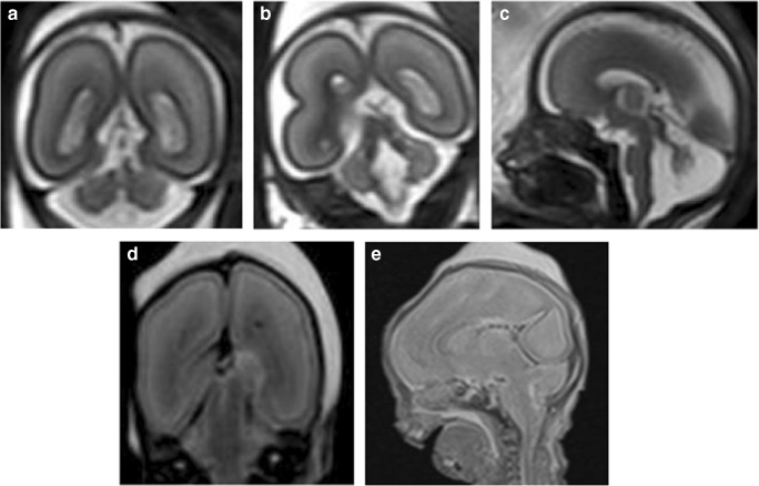
Fig. 6.
Abnormal cerebellar features in a 23-week gestation fetus. a, b Coronal T2-weighted iuMR images reveal features suggestive of an absent cerebellar vermis. The ‘buttocks sign’ is show in image a, and in image b, the ‘molar tooth appearance’ of elongated superior cerebellar peduncles is featured. c Sagittal T2-weighted iuMR image depicts the characteristic ‘figure of 7’ appearance of the elongated superior cerebellar peduncles and small cerebellum. d, e Coronal and sagittal T2-weighted PMMR images do not demonstrate the cerebellar abnormalities

