Abstract
Patient: Female, 63
Final Diagnosis: Recurrent desmoid tumor
Symptoms: Abdominal discomfort • abdominal fullness
Medication: —
Clinical Procedure: Abdominoplasty
Specialty: Plastic Surgery
Objective:
Rare co-existance of disease or pathology
Background:
Desmoid tumors are fibrous neoplasms that originate from the musculoaponeurotic structures in the body. Abdominal wall desmoid tumors are rare, but they can be locally aggressive, with high incidence of recurrence. These tumors are more common in young, fertile women. They frequently occur during or after pregnancy.
Case Report:
We present the case of a 63-year-old post-menopausal woman with a desmoid tumor of the anterior abdominal wall. She had no relevant family history. During abdominoplasty, an incidental mass was excised and biopsied, and was identified as a desmoid tumor with free margins. One year later, the patient presented with vague abdominal discomfort and feeling of heaviness. An incision was made through the previous abdominoplasty scar to maintain the aesthetic outcome. A large mass, arising from the abdominal wall and extending intra-abdominally, was excised and was determined to be a recurrent desmoid tumor.
Conclusions:
Recurrent anterior abdominal wall desmoid tumors in post-menopausal women are rare and locally aggressive, with a high risk of recurrence. During abdominal wall repair in abdominoplasty, desmoid tumor filaments might seed deep intra-abdominally. Therefore, it is necessary to take adequate safe margins before abdominal wall repair. Post-operatively, surgeons should keep a high index of suspicion for tumor recurrence.
MeSH Keywords: Abdominoplasty; Fibromatosis, Aggressive; Neoplasm Recurrence, Local
Background
Desmoid tumors are fibrous neoplasms that originate from the musculoaponeurotic structures in the body. In 1838, Muller first coined the term “desmoid”, which is derived from the Greek word “desmos”, which means tendon-like [1]. They contribute to around 3% of all soft tissue tumors and 0.03% of all neoplasms [2]. Despite their aggressive local infiltration, these tumors lack potential of distant metastasis [3]. They are most common in women of fertile age and are frequently found in the age group of 25–40 years. The most common site associated with these tumors is the anterior abdominal wall, with an incidence rate estimated at 50% [4].
Case Report
A 63-year-old post-menopausal woman, para 8, presented to our plastic surgery clinic seeking a body-contouring surgery for abdominal laxity. An abdominoplasty surgery was planned for excising and re-draping the redundant abdominal skin, abdominal folds, and addressing divarication of recti. On examination there was no palpable abdominal mass and the patient did not undergo any radiological investigations preoperatively.
During abdominoplasty, an incidental finding of a solid mass in the abdominal wall was made. It was an organized firm structure arising from the midline of the rectus sheath, in the para-umbilical region, both superior and inferior to the umbilicus, measuring 3×2 cm. It was excised in its entirety with no gross involvement of margins. The umbilicus was sacrificed and excised with the tumor, and neoumbilical reconstruction was done. The abdominal wall defect was closed primarily, and rectus sheath plication was performed. A histopathology study confirmed a desmoid tumor with free margins. Upon reviewing the patient’s history, she had no family history of desmoid tumors, familial adenomatous polyposis syndrome (FAP), or Gardener syndrome. After discharge, the patient was followed in the clinic regularly. She had a good aesthetic result and she was satisfied with the outcome.
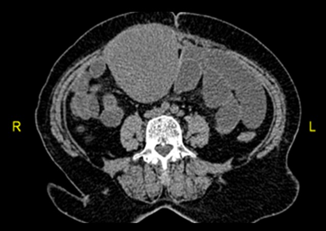
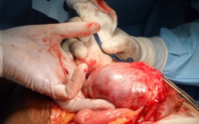
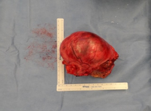
However, at 1 year after abdominoplasty, the patient presented with vague complains; she had a feeling of heaviness and discomfort in her abdomen. An abdominal examination was un-remarkable. A CT scan was performed, which showed a large abdominal mass (8×9×4 cm) arising from the abdominal wall and extending intra-abdominally (Figure 1). A second surgery was planned in collaboration with a general surgery team. The previous abdominoplasty scar was re-incised to give a better aesthetic outcome. A solid mass was found arising from the repaired abdominal wall, with an intra-abdominal extension that was adherent to the small bowel but easily dissectible from the tumor (Figures 2, 3).
Figure 1.

CT scan shows intra-abdominal extension of the desmoid tumor, which is adherent to the abdominal bowel.
Figure 2.

Excision of the desmoid tumor after secondary resection with intra-abdominal dissection.
Figure 3.

The dimensions of the tumor after excision.
A wide local excision with a 3-cm free margin of healthy tissue was confirmed by frozen section before the abdominal wall defect was reconstructed with a non-absorbable polypropylene surgical mesh (Surgipro, Covidien, Mansfield, MA, USA). Post-operatively, the patient had an uneventful recovery. After 7 days stay in the hospital, she was discharged. The histopathology report confirmed the diagnosis of desmoid tumor with free margins. Regular follow-ups in the clinic were scheduled.
Adjuvant chemotherapy was not recommended by the oncology team as the specimen had free margins. However, an MRI was performed at 6 months postoperatively, which did not show any residual or recurrent tumor. The patient was followed in the outpatient clinic for 2 years, during which she had full recovery. She had no functional deficit or abdominal hernia.
Discussion
Desmoid tumors are benign myofibroblastic neoplasms. Biologically, they show an intermediate behavior, which lies between benign fibrous lesions and fibrosarcomas [1]. They originate from the muscle aponeurosis and are classified as deep fibromatoses [2,4].
These tumors are commonly associated with fertile women during or after their pregnancy. It has been shown that estrogen has a proliferative effect on the fibroblasts. Some patients have shown regression after reaching menopause [4]. The association of abdominal and pelvic surgery is also well established with desmoid tumors, as well as an association with trauma, estrogen therapy, FAP, and Gardner syndrome [5–10].
Desmoid tumors are divided into 5 subgroups: extra-abdominal, intra-abdominal, multiple, multiple familial, and as part of Gardner’s syndrome. Abdominal wall desmoid tumors arise from musculoaponeurotic structures. The rectus muscle and internal oblique muscle and their fascial coverings are most frequently affected. However, tumors originating from the external oblique muscle and the transversalis muscle or fascia are less common [11].
The aim for surgical management of desmoid tumors should be radical resection with free margins, which might leave soft tissue defects, as in our case. However, complete surgical excision is the most effective treatment to will reduce the risk of local recurrence. Adjuvant treatment, including chemotherapy and repeat surgery, may be required in excision of extensive cases [12,13]. Open surgical resection has been the recognized treatment for such tumors. Recently, laparoscopic techniques have been reported, which may become more established in the future if promising results and no increase in recurrence is reported [14]. However, we selected a more conventional approach in the second surgery since it was a recurrent tumor with abdominal wall origin after abdominoplasty surgery.
Desmoid tumors have a high chance of recurrence; it was reported to be as high as 70%. A positive surgical margin is a major risk factor for recurrence [15,16]. Therefore, surgeons should keep a high index of suspicion for possible tumor recurrence. They are known to be benign tumors, but can be locally aggressive [17]. They may cause intestinal obstruction or fistulization. For these reasons, aggressive treatment strategies were historically employed to completely extirpate disease. Individuals with Gardner syndrome have an increased incidence of abdominal desmoids that are present in the abdominal wall, mesentery, or retro-peritoneum [4,18].
In case of recurrent abdominal desmoid tumors, a radical re-section with intra-operative margin evaluation by frozen section, followed by immediate mesh reconstruction, may be a safe and effective procedure [19]. Follow-up with MRI is recommended for early detection of recurrence. However, there is no general consensus in the literature regarding the best MRI time during follow-up or the required frequency.
Conclusions
Recurrent anterior abdominal wall desmoid tumors in post-menopausal women is rare and locally aggressive, with a high risk of recurrence. During abdominal wall repair in abdominoplasty, desmoid tumor filaments might seed deep intra-abdominally. Therefore, it is necessary to take enough safe margins before abdominal wall repair. Post-operatively, surgeons should keep a high index of suspicion for tumor recurrence.
References:
- 1.Shields CJ, Winter DC, Kirwan WO, Redmond HP. Review desmoid tumors. Eur J Surg Oncol. 2001;27(8):701–6. doi: 10.1053/ejso.2001.1169. [DOI] [PubMed] [Google Scholar]
- 2.Fletcher CDM. Myofibroblastic tumors: An update. Verh Dtsch Ges Path. 1998;82:75–82. [PubMed] [Google Scholar]
- 3.Kiel KD, Suit HD. Radiation therapy in the treatment of aggressive fibromatoses (desmoid tumors) Cancer. 1984;54:2051–55. doi: 10.1002/1097-0142(19841115)54:10<2051::aid-cncr2820541002>3.0.co;2-2. [DOI] [PubMed] [Google Scholar]
- 4.Economou A, Pitta X, Andreadis E, et al. Desmoid tumor of the abdominal wall: A case report. J Med Case Rep. 2011;5:326. doi: 10.1186/1752-1947-5-326. [DOI] [PMC free article] [PubMed] [Google Scholar]
- 5.Crago AM, Denton B, Salas S, et al. A prognostic nomogram for prediction of recurrence in desmoid fibromatosis. Ann Surg. 2013;258(2):347–53. doi: 10.1097/SLA.0b013e31828c8a30. [DOI] [PMC free article] [PubMed] [Google Scholar]
- 6.Teo HEL, Peh WCG, Shek TWH. Case 84: The desmoid tumor of the abdominal wall. Radiology. 2005;236:81–84. doi: 10.1148/radiol.2361031038. [DOI] [PubMed] [Google Scholar]
- 7.Kumar V, Khanna S, Khanna AK, Khanna R. Desmoid tumors: The experience of 32 cases and review of the literature. Indian J Cancer. 2009;46:34–39. doi: 10.4103/0019-509x.48593. [DOI] [PubMed] [Google Scholar]
- 8.Lahat G, Nachmany I, Itzkowitz E, et al. Surgery for sporadic abdominal desmoid tumor: Is low/no recurrence an achievable goal? IMAJ. 2009;11:398–402. [PubMed] [Google Scholar]
- 9.Overhaus M, Decker P, Fischer HP, et al. Desmoid tumors of the abdominal wall: A case report. World J Surg Oncol. 2003;1(1):11. doi: 10.1186/1477-7819-1-11. [DOI] [PMC free article] [PubMed] [Google Scholar]
- 10.Efthimiopoulos GA, Chatzifotiou D, Drogouti M, Zafiriou G. Primary asymptomatic desmoid tumor of the mesentery. Am J Case Rep. 2015;16:160–63. doi: 10.12659/AJCR.892521. [DOI] [PMC free article] [PubMed] [Google Scholar]
- 11.Casillas J, Sais GJ, Greve JL, et al. Imaging of intra and extra-abdominal desmoid tumors. Radiographics. 1991;11:959–68. doi: 10.1148/radiographics.11.6.1749859. [DOI] [PubMed] [Google Scholar]
- 12.Ramirez RN, Otsuka NY, Apel DM, Bowen RE. Desmoid tumor in the pediatric population: A report of two cases. J Pediatr Orthop B. 2009;18(3):141–44. doi: 10.1097/BPB.0b013e3283298923. [DOI] [PubMed] [Google Scholar]
- 13.Bertani E, Chiappa A, Testori A, et al. Desmoid tumors of the anterior abdominal wall: Results from a monocentric surgical experience and review of the literature. Ann Surg Oncol. 2009;16(6):1642–49. doi: 10.1245/s10434-009-0439-z. [DOI] [PubMed] [Google Scholar]
- 14.Meshikhes AW, Al-Zahrani H, Ewies T. Laparoscopic excision of abdominal wall desmoid tumor. Asian J Endosc Surg. 2016;9(1):79–82. doi: 10.1111/ases.12257. [DOI] [PubMed] [Google Scholar]
- 15.Huang PW, Tzen CY. Prognostic factors in desmoid-type fibromatosis: A clinicopathological and immunohistochemical analysis of 46 cases. Pathology. 2010;42(2):147–50. doi: 10.3109/00313020903494078. [DOI] [PubMed] [Google Scholar]
- 16.Meazza C, Bisogno G, Gronchi A, et al. Aggressive fibromatosis in children and adolescents: the Italian experience. Cancer. 2010;116(1):233–40. doi: 10.1002/cncr.24679. [DOI] [PubMed] [Google Scholar]
- 17.Lewis JJ, Boland PJ, Leung DH, et al. The enigma of desmoid tumors. Ann Surg. 1999;229(6):866–72. doi: 10.1097/00000658-199906000-00014. discussion 872–73. [DOI] [PMC free article] [PubMed] [Google Scholar]
- 18.Palladino E, Nsenda J, Siboni R, Lechner C. A giant mesenteric desmoid tumor revealed by acute pulmonary embolism due to compression of the inferior vena cava. Am J Case Rep. 2014;15:374–77. doi: 10.12659/AJCR.891044. [DOI] [PMC free article] [PubMed] [Google Scholar]
- 19.Bhama PK, Chugh R, Baker LH, Doherty GM. Gardner’s syndrome in a 40-year-old woman: Successful treatment of locally aggressive desmoid tumors with cytotoxic chemotherapy. World J Surg Oncol. 2006;4:96. doi: 10.1186/1477-7819-4-96. [DOI] [PMC free article] [PubMed] [Google Scholar]


