Abstract
Objectives:
To assess phosphate and osmolarity levels of chronically administered eye drops commercially available in Turkey.
Materials and Methods:
A total of 53 topical eye drops including 18 antiglaucoma drugs, 4 nonsteroidal anti-inflammatory drugs (NSAIDs), 10 corticosteroids, 7 antihistaminics, and 14 artificial tears identified using the Vademecum Modern Medications Guideline (2018) were included in the study. Phosphate levels were assessed using Roche Cobas C501 analyzer (Roche Diagnostics GmbH, Mannheim, Germany) and the respective kits. Osmolarity was assessed using Vescor Vapro 5600 vapor pressure osmometer (Sanova Medical Systems, Vienna, Austria). Mean phosphate and osmolarity levels were obtained after averaging three measurements. Eye drops were categorized as isoosmolar, hypoosmolar and hyperosmolar based on physiologic tear osmolarity range (296.5±9.8 mOsm/L).
Results:
The highest phosphate concentration was found in the antiglaucoma group (20.3±35.4 mmol/L), followed by antihistaminics (17.3±17.9 mmol/L), corticosteroids (15.2±19.1 mmol/L), artificial tears (0.8±1.0), and NSAIDs (0.04±0.08). Percentage of medications in the hyperosmolar category was highest in the NSAI group (75%), followed by antihistaminics (43%), corticosteroids (20%), and antiglaucoma drugs (19%). Nearly all of the artificial tear formulations were in the hypoosmolar (71%) or isoosmolar (21%) categories.
Conclusion:
Approximately 40% of glaucoma medications and approximately 60% of corticosteroid and antihistaminic medications had a phosphate concentration higher than the physiologic tear phosphate level (1.45 mmol/L).
Keywords: Phosphate, eye drops, osmolarity, corneal calcification
Introduction
While topical eye drops have an important place in the treatment of eye diseases, long-term and inappropriate use may cause serious complications and side effects affecting the ocular surface. These side effects may be caused by an active pharmaceutical ingredient, preservative, or vehicle in the topical formulation.1 The side effect profiles of active ingredients are thoroughly investigated during the stages of drug development, and the process of monitoring for adverse effects also continues after the molecule enters the market. After recent studies revealed that preservatives can also cause severe toxicity, efforts have been made to develop less toxic preservative molecules or preservative-free eye drops. However, the potential toxicity of molecules comprising eye drop vehicles has been a relatively neglected topic that has not been given due importance.
Vehicles are involved in buffering eye drops and ensure that the formulation has the appropriate tonicity and viscosity.#*#ref2#*# Buffering agents include molecules like acetic, boric, and hydrochloric acid, potassium or sodium bicarbonate, phosphate, and citrate.1 Phosphate, a commonly used buffer, is a vehicle with high buffering capacity that stabilizes the pH level at 7.4, and can also be found in some formulations as part of the active ingredient.1,3,4 In addition, it has the added advantage of making corticosteroid-containing solutions more transparent.3
Although phosphate is an effective buffer, it interacts with calcium cations on the ocular surface to disrupt the structure of the precorneal tear film and form insoluble hydroxyapatite [Ca5(PO4)3OH] or calcium phosphate crystals in the cornea.2,5,6,7 The resulting crystals cause irreversible stromal opacification and reduced vision, and can also have a serious impact on patient comfort.5,6,8 An example of this crystallization was previously demonstrated in a patient with chemical burn of the ocular surface that was irrigated with a phosphate-buffered saline solution.9 The development of irreversible corneal calcification after the use of phosphate-buffered artificial tears for ocular surface disorders occurs for a similar reason.5 The extent of accumulation depends on factors such as the size of the epithelial defect, the presence of dry eye, the pH and tonicity of the formulation, and the frequency and duration of use.2,9,10
The aim of our study was to examine the phosphate concentrations and osmolarity levels of chronically administered eye drops commercially available in Turkey. We hereby intend to highlight the distinct importance of phosphate levels in eye drops in addition to the known hazards imposed by the active ingredients and preservatives.
Materials and Methods
The Vademecum Modern Drug Directory (2018) was screened for antiglaucoma drugs, nonsteroidal anti-inflammatory drugs (NSAIDs), corticosteroids, antihistamines, and artificial tears for chronic topical use that are commercially available in Turkey. A total of 53 topical drugs, including 18 antiglaucoma drugs, 4 NSAIDs, 10 corticosteroids, 7 antihistamines, and 14 artificial tears, were included in the study in order to examine their phosphate and osmolarity levels (Table 1). Because this study did not involve humans or the use of human biological material, it was considered exempt from ethics board approval by the Ethics Committee of Eskişehir Osmangazi University. Topical formulations with high viscosity were excluded from the research due to technical reasons. Phosphate levels were determined at the Medical Biochemistry Department Laboratory of the Medical Faculty at Eskişehir Osmangazi University using a Roche Cobas C501 analyzer (Roche Diagnostics GmbH, Mannheim, Germany) with an inorganic phosphate kit based on the molybdate UV method.11 The kit has a measurement range of 0.1-6.46 mmol/L and a lower limit of detection of 0.1 mmol/L. Samples above the measurable range were diluted and analyzed again. The kit has good reproducibility (CV<1.5%). Osmolarity of the topical drops was evaluated with a Vescor Vapro 5600 model steam pressure osmometer (Sanova Medical Systems, Vienna, Austria) found in the same laboratory. Three levels of control were used to calibrate the device: low (100±2 mOsm/L), normal (290±3 mOsm/L), and high (1000±5 mOsm/L). The phosphate and osmolarity levels of each eye drop were determined three times and the average values were included in the analysis.
Table 1. Phosphate and osmolarity levels in the different categories of eye drops.
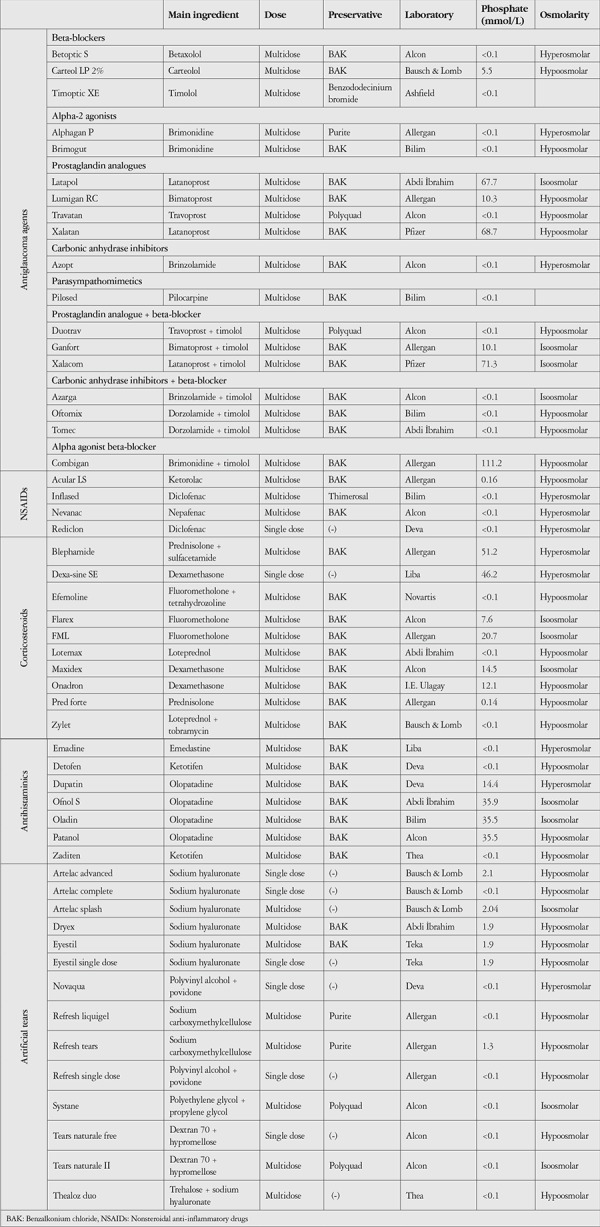
Information about the preservatives found in the drops was obtained from the Vademecum Modern Drug Directory.
Drops within the physiological osmolarity range of tears (296.5±9.8 mOsm/L)12 were classified as isoosmolar, and those below and above this range were classified as hypoosmolar and hyperosmolar, respectively.
Based on their phosphate concentrations, the topical drops were classified as being within physiological range (≤1.45 mmol/L), slightly high (1.45-25 mmol/L), moderately high (25-50 mmol/L), and very high (≥50 mmol/L).2
Statistical Analyses
All statistical analyses were made with SPSS version 21.0 (SPSS, Inc. IBM, Chicago, IL). The average phosphate values of drugs with different preservative ingredients and in the different osmolarity categories were evaluated with Kruskal-Wallis test. Statistical significance was set at p<0.05.
Results
The phosphate concentrations and osmolarity categories of the eye drops included in the study are summarized in Table 1. The highest measured average phosphate level was in the antiglaucoma group (20.3±35.4 mmol/L), followed by antihistamines (17.3±17.9 mmol/L), corticosteroids (15.2±19.1 mmol/L), artificial tears (0.8±1.0 mmol/L), and NSAIDs (0.04±0.08 mmol/L) (Figure 1). Thirty-one (58.5%) of the 53 topical drops contained phosphate levels within the physiological range. Preparations containing moderately and very high levels of phosphate accounted for 22.2% of the antiglaucoma drops and 42.9% of the antihistamines (Figure 2).
Figure 1.
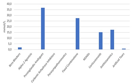
Mean phosphate levels of different drug groups
NSAIDs: Nonsteroidal anti-inflammatory drugs
Figure 2.
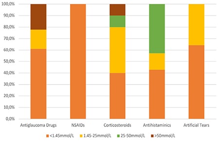
Percent distribution of phosphate levels in the different drug groups
NSAIDs: Nonsteroidal anti-inflammatory drugs
In the antiglaucoma group, it was noted that drops containing latanoprost contained especially high phosphate levels (Table 1). In the antihistamine group, drops containing olopatadine were found to contain high levels of phosphate, while other drops contained trace amounts of phosphate (Table 1). In the artificial tear group, most preparations had trace amounts of phosphate, while those containing sodium hyaluronate had slightly high levels of phosphate (Table 1).
When different drug groups were evaluated based on their osmolarity levels, it was found that preparations in the NSAID group were of hyperosmolar character, while preparations in the artificial tear group were mostly hypoosmolar or isoosmolar (Figure 3).
Figure 3.
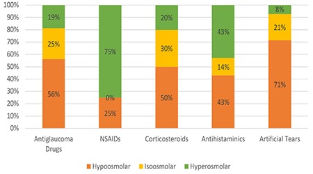
Percent distribution of osmolarity levels in the different drug groups
NSAIDs: Nonsteroidal anti-inflammatory drugs
Evaluation of the phosphate levels of drugs in different osmolarity categories showed that hypoosmolar and hyperosmolar drugs contained similar levels of phosphate (9.0±24.6 mmol/L and 10.2±19.6 mmol/L, respectively); isoosmolar drugs had a relatively higher mean phosphate level (22.1±25.6 mmol/L), but the difference was not statistically significant (p>0.05) (Figure 4).
Figure 4.
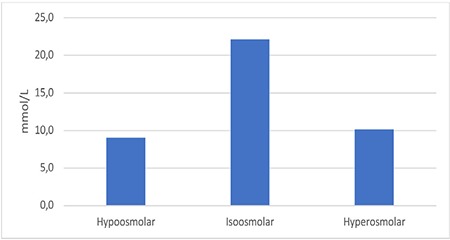
Average phosphate levels of drugs by osmolarity category
It was noted that of the 53 drugs, 34 contained benzalkonium chloride (BAK) as a preservative, 9 contained a non-BAK preservative, and the remaining 10 drugs contained no preservatives. When phosphate levels were evaluated based on preservative, the highest phosphate level was in those containing BAK (16.9±27.4 mmol/L), followed by preservative-free drops (5.2±14.4 mmol/L) and drops containing non-BAK preservatives (0.14±0.42 mmol/L) (Figure 5).
Figure 5.
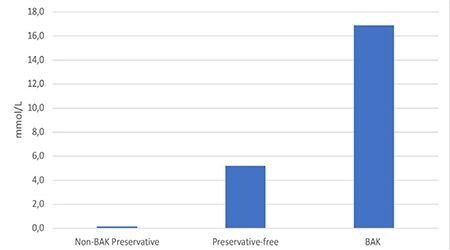
Average phosphate levels according to preservative
BAK: Benzalkonium chloride
There was a significant difference between the group containing BAK and the group containing non-BAK preservatives (p=0.04), but the other comparisons did not yield statistically significant results.
Discussion
Corneal calcification can result in severely reduced vision and in most cases irreversible corneal opacification, and may be associated with long-term use of eye drops with high phosphate content.5,6,13 The formation of crystals without visible calcification due to the use of high-viscosity artificial tears can also lead to irritation and thereby disrupt patient comfort.8 Rapid corneal calcification has also been reported in patients with large epithelial defects that were irrigated with phosphate-buffered solutions after chemical burns.9,14,15 In this study, we found that 22 (41.5%) of the 53 drops analyzed contained levels of phosphate exceeding physiological concentration (0-1.34 mmol/L) and that the majority of these were in the antiglaucoma and antihistamine drug groups.
The deposition of hydroxyapatite [Ca5(PO4)3OH] crystals in Bowman’s layer and the superficial stroma of the cornea is called band keratopathy.16 In cases with both epithelial damage and disruption of Bowman’s membrane, accumulation occurs in the deeper corneal stroma and Descemet’s membrane in the form of calcareous degeneration.17 The solubility of these crystals decreases with alkaline pH and high temperature.18 The pH value (physiological range 7.6-7.8)19 and tonicity of the ocular surface, extent of epithelial damage, and presence of inflammation and barrier dysfunction are among the factors responsible for the development of corneal calcification.2,9 Especially in dry eye patients, the tear film becomes more alkaline and hyperosmolar.20,21,22 An alkaline shift in the tear film has also been reported in association with age, independent of dry eye disease.23,24 The hyperosmolar state that occurs in dry eye disease is known to trigger the release of inflammatory mediators and proteases, which cause epithelial destruction.25 Similarly, topical drops with a hyperosmolar character have also been shown to alter tear osmolarity and increase inflammation.26 As ocular inflammation is a known risk factor for corneal calcification, hyperosmolar drops are not recommended for patients with a predisposition to corneal calcification.27,28
Although the phosphate content of the artificial tears analyzed in our study were within physiological limits, factors such as high-frequency use, inadequate lacrimal drainage, extended tear turnover time, and the high viscosity of artificial tears can increase the duration of contact between the ocular surface and the phosphate found in the formulation and thus lead to a tendency for calcification.2,10 Because dry eye also involves an inflammatory component, treatment may involve the intermittent use of steroids. Full-thickness calcification in the corneal stroma following the long-term use of dexamethasone phosphate was reported in a patient with Stevens-Johnson syndrome (SJS).6 Therefore, in the presence of an epithelial defect, it may be beneficial to prefer topical steroids that have low phosphate content or contain a non-phosphate buffer and are preferably acidic. Notably, the only BAK-free formulation in the topical steroid group contains phosphate at a concentration above the physiological limit.
In our study, the highest phosphate concentrations were detected in the antiglaucoma drops. Drops containing latanoprost in particular contained approximately 50 times more phosphate than the upper limit of the physiological range. Although the more acidic pH values (≈6.4)10 of these drops may seem like an advantage, the high phosphate concentrations increase their risks. In contrast, although bimatoprost drops and bimatoprost fixed combination drops contain less phosphate, they are more alkaline.10 The disclaimers in the package inserts of both of these prostaglandin analogues stating that “in rare cases, patients with severe damage to the cornea may develop cloudy patches due to the calcium build-up during treatment” should be evaluated in this context.29,30 Considering that predisposition to phosphate deposition in the cornea is a pH-dependent process, it will be valuable to demonstrate the effect of both preparations in vivo. In addition to drug pH values, the presence of ocular surface inflammation in glaucoma patients may be a factor that increases the risk of corneal deposits. Although trace amounts of phosphate were detected in the drops containing timolol in our study, accumulation in the superficial corneal stroma associated with timolol has been reported in the literature.31,32
Combining the reduced tear film breakup time, ocular pH changes, and ocular surface temperature and chronic inflammation that occur in allergic conjunctivitis with the chronic use of drugs containing high phosphate levels may promote the formation of corneal deposits.33,34,35 It is important to evaluate the topical antihistamines, steroids, and artificial tears used in treatment with this in mind. Shield ulcers that may occur in vernal conjunctivitis are another condition in which the risk of corneal disposition should be assessed.
Conclusion
In summary, cases of acute or chronic corneal calcification associated with the use of topical drops or irrigation solutions with high phosphate levels have been reported in patients with chemical burns, dry eye, and chronic keratoconjunctivitis secondary to SJS.5,6,9 Considering evidence that phosphate-buffered tears but not citrate-buffered tears caused corneal calcification in some rabbits with mechanical abrasion-induced epithelial defect, drops containing a non-phosphate buffer can be considered for at-risk patients.18 A European Medicines Agency report evaluating 117 cases related to this topic emphasized that there is a possible association between corneal calcification and the use of topical drops in patients with corneal surface disorders.36 The reported concluded by stating that due to the very low risk, there is no need to refrain from using phosphate buffered drops, but that the risk-benefit balance should be considered when prescribing these drugs to patients with corneal damage.36 Knowing the chemical structure of topical formulations and selecting drops that have suitable tonicity and pH according to the disease profile and contain a buffer that will not promote accumulation will help prevent ocular surface complications associated with the use of eye drops.
Footnotes
Ethics
Ethics Committee Approval: Because this study did not involve humans or the use of human biological material, it was considered exempt from ethics board approval by the Ethics Committee of Eskişehir Osmangazi University.
Informed Consent: Received.
Peer-review: Externally and internally peer-reviewed.
Authorship Contributions
Concept: Nilgün Yıldırım, İbrahim Özkan Alataş, Eray Atalay, Design: Nilgün Yıldırım, İbrahim Özkan Alataş, Data Collection or Processing: Onur Özalp, Eray Atalay, Zeynep Küskü Kiraz, İbrahim Özkan Alataş, Nilgün Yıldırım, Analysis or Interpretation: Eray Atalay, Zeynep Küskü Kiraz, İbrahim Özkan Alataş, Nilgün Yıldırım, Literature Search: Onur Özalp, Eray Atalay, Nilgün Yıldırım, Writing: Onur Özalp, Eray Atalay, Nilgün Yıldırım.
Conflict of Interest: No conflict of interest was declared by the authors.
Financial Disclosure: The authors declared that this study received no financial support.
References
- 1.Fiscella RG. Ophthalmic Drug Formulations. In: Bartlett JD, Jaanus SD, eds. Clinical Ocular Pharmacology: Butterworth-Heinemann/Elsevier. 2008. [Google Scholar]
- 2.Bernauer W, Thiel MA, Langenauer UM, Rentsch KM. Phosphate concentration in artificial tears. Graefes Arch Clin Exp Ophthalmol. 2006;244:1010–1014. doi: 10.1007/s00417-005-0219-9. [DOI] [PubMed] [Google Scholar]
- 3.Polansky JR, Weinreb RN. Anti-inflammatory agents-steroids as antiinflammatory agents. In: Sears MI, ed. Handbook of Experimental Pharmacology. Berlin Heidelberg New York; Springer. 1984. [Google Scholar]
- 4.Bernauer W, Thiel MA, Rentsch KM. Phosphate concentration in ophthalmic corticoid preparations. Graefes Arch Clin Exp Ophthalmol. 2008;246:975–978. doi: 10.1007/s00417-008-0788-5. [DOI] [PubMed] [Google Scholar]
- 5.Bernauer W, Thiel MA, Kurrer M, Heiligenhaus A, Rentsch KM, Schmitt A, Heinz C, Yanar A. Corneal calcification following intensified treatment with sodium hyaluronate artificial tears. Br J Ophthalmol. 2006;90:285–288. doi: 10.1136/bjo.2005.082792. [DOI] [PMC free article] [PubMed] [Google Scholar]
- 6.Schlotzer-Schrehardt U, Zagorski Z, Holbach LM, Hofmann-Rummelt C, Naumann GO. Corneal stromal calcification after topical steroid-phosphate therapy. Arch Ophthalmol. 1999;117:1414–1418. doi: 10.1001/archopht.117.10.1414. [DOI] [PubMed] [Google Scholar]
- 7.Popiela MZ, Hawksworth N. Corneal calcification and phosphates: do you need to prescribe phosphate free? J Ocul Pharmacol Ther. 2014;30:800–802. doi: 10.1089/jop.2014.0054. [DOI] [PubMed] [Google Scholar]
- 8.Ridder WH, Lamotte JO, Ngo L, Fermin J. Short-term effects of artificial tears on visual performance in normal subjects. Optom Vis Sci. 2005;82:370–377. doi: 10.1097/01.opx.0000162646.30666.e3. [DOI] [PubMed] [Google Scholar]
- 9.Daly M, Tuft SJ, Munro PM. Acute corneal calcification following chemical injury. Cornea. 2005;24:761–765. doi: 10.1097/01.ico.0000154040.80442.8b. [DOI] [PubMed] [Google Scholar]
- 10.Martinez-Soroa I, de Frutos-Lezaun M, Ostra Beldarrain M, Egia Zurutuza A, Irastorza Larburu MB, Bachiller Cacho MP. Determination of phosphate concentration in glaucoma eye drops commercially available in Spain. Arch Soc Esp Oftalmol. 2016;91:363–371. doi: 10.1016/j.oftal.2016.02.010. [DOI] [PubMed] [Google Scholar]
- 11.Henry RJ. Clinical Chemistry: Principles and Technics. In: Henry RJ, ed. 2nd ed. New York; Harper & Row. 1974. [Google Scholar]
- 12.Versura P, Profazio V, Campos EC. Performance of tear osmolarity compared to previous diagnostic tests for dry eye diseases. Curr Eye Res. 2010;35:553–564. doi: 10.3109/02713683.2010.484557. [DOI] [PubMed] [Google Scholar]
- 13.Schrage NF, Kompa S, Ballmann B, Reim M, Langefeld S. Relationship of eye burns with calcifications of the cornea? Graefes Arch Clin Exp Ophthalmol. 2005;243:780–784. doi: 10.1007/s00417-004-1089-2. [DOI] [PubMed] [Google Scholar]
- 14.Schrage NF, Schlossmacher B, Aschenbernner W, Langefeld S. Phosphate buffer in alkali eye burns as an inducer of experimental corneal calcification. Burns. 2001;27:459–464. doi: 10.1016/s0305-4179(00)00148-0. [DOI] [PubMed] [Google Scholar]
- 15.Huang Y, Meek KM, Mangat H, Paterson CA. Acute calcification in alkaliinjured rabbit cornea treated with synthetic inhibitor of metalloproteinases (SIMP) Cornea. 1998;17:423–432. doi: 10.1097/00003226-199807000-00014. [DOI] [PubMed] [Google Scholar]
- 16.Hinzpeter EN, Naumann GOH. Cornea and sclera. In: Naumann GOH, Apple DJ, eds. Pathology of the eye New York; Springer-Verlag. 1986. [Google Scholar]
- 17.Lake D, Tarn A, Ayliffe W. Deep corneal calcification associated with preservative-free eyedrops and persistent epithelial defects. Cornea. 2008;27:292–296. doi: 10.1097/ICO.0b013e31815c5a24. [DOI] [PubMed] [Google Scholar]
- 18.Schrage NF, Frentz M, Reim M. Changing the composition of buffered eyedrops prevents undesired side effects. Br J Ophthalmol. 2010;94:1519–1522. doi: 10.1136/bjo.2009.177386. [DOI] [PubMed] [Google Scholar]
- 19.Chen FS, Maurice DM. The pH in the precorneal tear film and under a contact lens measured with a fluorescent probe. Exp Eye Res. 1990;50:251–259. doi: 10.1016/0014-4835(90)90209-d. [DOI] [PubMed] [Google Scholar]
- 20.Norn MS. Tear fluid pH in normals, contact lens wearers, and pathological cases. Acta Ophthalmol (Copenh). 1988;66:485–489. doi: 10.1111/j.1755-3768.1988.tb04368.x. [DOI] [PubMed] [Google Scholar]
- 21.Khurana AK, Chaudhary R, Ahluwalia BK, Gupta S. Tear film profile in dry eye. Acta Ophthalmol (Copenh). 1991;69:79–86. doi: 10.1111/j.1755-3768.1991.tb01997.x. [DOI] [PubMed] [Google Scholar]
- 22.Willcox MDP, Argueso P, Georgiev GA, Holopainen JM, Laurie GW, Millar TJ, Papas EB, Rolland JP, Schmidt TA, Stahl U, Suarez T, Subbaraman LN, Ucakhan OÖ, Jones L. TFOS DEWS II Tear Film Report. Ocul Surf. 2017;15:366–403. doi: 10.1016/j.jtos.2017.03.006. [DOI] [PMC free article] [PubMed] [Google Scholar]
- 23.Coles WH, Jaros PA. Dynamics of ocular surface pH. Br J Ophthalmol. 1984;68:549–552. doi: 10.1136/bjo.68.8.549. [DOI] [PMC free article] [PubMed] [Google Scholar]
- 24.Andres S, Garcia ML, Espina M, Valero J, Valls O. Tear pH, air pollution, and contact lenses. Am J Optom Physiol Opt. 1988;65:627–631. doi: 10.1097/00006324-198808000-00006. [DOI] [PubMed] [Google Scholar]
- 25.Craig JP, Nelson JD, Azar DT, Belmonte C, Bron AJ, Chauhan SK, de Paiva CS, Gomes JAP, Hammitt KM, Jones L, Nichols JJ, Nichols KK, Novack GD, Stapleton FJ, Willcox MDP, Wolffsohn JS, Sullivan DA. TFOS DEWS II Report Executive Summary. Ocul Surf. 2017;15:802–812. doi: 10.1016/j.jtos.2017.08.003. [DOI] [PubMed] [Google Scholar]
- 26.Dutescu RM, Panfil C, Schrage N. Osmolarity of prevalent eye drops, side effects, and therapeutic approaches. Cornea. 2015;34:560–566. doi: 10.1097/ICO.0000000000000368. [DOI] [PubMed] [Google Scholar]
- 27.Troiano P, Monaco G. Effect of hypotonic 0.4% hyaluronic acid drops in dry eye patients: a cross-over study. Cornea. 2008;27:1126–1130. doi: 10.1097/ICO.0b013e318180e55c. [DOI] [PubMed] [Google Scholar]
- 28.Aragona P, Di Stefano G, Ferreri F, Spinella R, Stilo A. Sodium hyaluronate eye drops of different osmolarity for the treatment of dry eye in Sjögren’s syndrome patients. Br J Ophthalmol. 2002;86:879–884. doi: 10.1136/bjo.86.8.879. [DOI] [PMC free article] [PubMed] [Google Scholar]
- 29.No authors listed. XALATAN® % 0.005 Göz Damlası Kullanma Talimatı. Pfizer [Google Scholar]
- 30.No authors listed. LUMİGAN RC %0.01 Göz Damlası Kullanma Talimatı. Allergan [Google Scholar]
- 31.Huige WM, Beekhuis WH, Rijneveld WJ, Schrage N, Remeijer L. Unusual deposits in the superficial corneal stroma following combined use of topical corticosteroid and beta-blocking medication. Doc Ophthalmol. 1991;78:169–175. doi: 10.1007/BF00165677. [DOI] [PubMed] [Google Scholar]
- 32.Huige WM, Beekhuis WH, Rijneveld WJ, Schrage N, Remeijer L. Deposits in the superficial corneal stroma after combined topical corticosteroid and beta-blocking medication. Eur J Ophthalmol. 1991;1:198–199. doi: 10.1177/112067219100100407. [DOI] [PubMed] [Google Scholar]
- 33.Toda I, Shimazaki J, Tsubota K. Dry eye with only decreased tear break-up time is sometimes associated with allergic conjunctivitis. Ophthalmology. 1995;102:302–309. doi: 10.1016/s0161-6420(95)31024-x. [DOI] [PubMed] [Google Scholar]
- 34.Hara Y, Shiraishi A, Yamaguchi M, Kawasaki S, Uno T, Ohashi Y. Evaluation of allergic conjunctivitis by thermography. Ophthalmic Res. 2014;51:161–166. doi: 10.1159/000357105. [DOI] [PubMed] [Google Scholar]
- 35.Ichihashi Y, Ide T, Kaido M, Ishida R, Hatou S, Tsubota K. Short breakup time type dry eye has potential ocular surface abnormalities. Taiwan J Ophthalmol. 2015;5:68–71. doi: 10.1016/j.tjo.2015.02.004. [DOI] [PMC free article] [PubMed] [Google Scholar]
- 36.No authors listed. Committee for Medicinal Products for Human Use (CHMP) Questions and answers on the use of phosphates in eye drops. European Medicines Agency. 2012. [Google Scholar]


