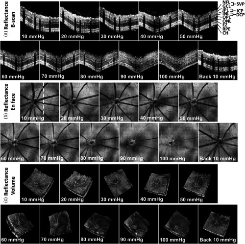
Fig. 2.
Reflectivity of the retinal NFL is reduced with acute IOP elevation, as demonstrated by sequential darkening in (a) B-scans, (b) en face images (), and (c) volumetric visualization. Each B-scan was averaged by three repeated frames. The white dashed line in (b) indicates the position of B-scans in (a). NFL, nerve fiber layer; IPL, inner plexiform layer; INL, inner nuclear layer; OPL, outer plexiform layer; ONL, outer nuclear layer; EZ, ellipsoid zone; RPE, retinal pigment epithelium; CH, choroid; SVP, superficial vascular plexus; ICP, intermediate capillary plexus; and DCP, deep capillary plexus.

