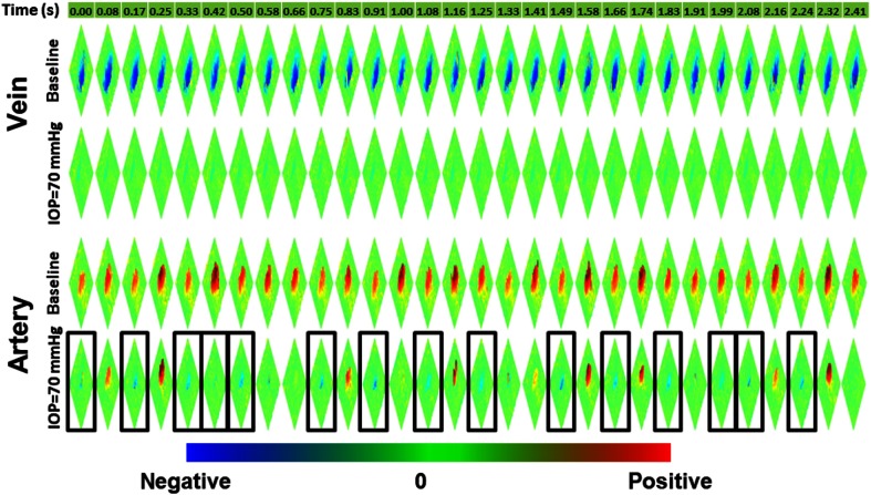
Fig. 5.
Arterial flow reversal at shown by a series of Doppler OCT (30 frames within 2.4 s) of a representative artery, compared to the constant flow direction in a vein. Doppler OCT images with arterial flow reversal are annotated with black boxes. Red: positive blood flow. Blue: negative blood flow.

