

Abstract
Aim
To evaluate the effect of cold and room-temperature normal saline as a final irrigation on post-endodontic pain and to compare the post-endodontic pain level between the different protocols.
Materials and methods
A randomized controlled trial was conducted on 105 patients who underwent RCT and were distributed blindly and randomly into three equal groups (n = 35): Group 1, Cryotherapy group; Group 2, Room- temperature normal saline group; and Group 3, Control group. The patients were asked to fill out the VAS questionnaire and register their post-endodontic pain at 6, 24, and 48 h. One-way analysis of variance (ANOVA) and was used to determine statistical difference (α = 0.05) among the groups. Tuckey’s test was used to calculate P-value (α = 0.05) between two each group.
Results
The highest post-endodontic pain was in Group 3. There was no significance difference between Group 1 and Group 2.
Conclusion
Final flushing of the canal with saline either cold or at room temperature was effective for post-endodontic pain control. This can be promising as an essential step in endodontic treatment to reduce post-endodontic pain. The room-temperature saline as final irrigation showed comparable results to intra-canal cryotherapy.
Keywords: Cryotherapy, Endodontics, Post-operative pain, Single-visit RCT
1. Introduction
Prevalence of post- endodontic pain was high but decreased within one day after root canal treatment (RCT) and reached a minimal level within seven days (Pak and White, 2011). 47–60% of cases with asymptomatic pulpitis that advanced to pulp necrosis had a higher chance to develop post-endodontic pain, although the pulp state effect on post-endodontic pain is controversial (Sipavičiūtė and Manelienė, 2014).
The most common factor cause the post-endodontic is remnants of microorganisms (AlRahabi, 2017). Endodontic irrigation, gutta percha, endodontic sealer, and intra = canal medication are all factors that can cause irritation and post-endodontic pain (Siqueira and Barnett, 2004, AlRahabi, 2017). There is no difference between single-visit and multiple-visit RCT on post-endodontic pain (Manfredi et al., 2016, AlRahabi, 2017).
According to a recent systematic review and meta-analysis of nonsteroidal anti-inflammatory drugs (NSAIDS) for management of post-endodontic pain, ibuprofen is an effective analgesic drug to control post-endodontic pain (Smith et al., 2017).
Cryotherapy is the application of cold material to lower the temperature of the surrounding tissues to control pain (Nadler et al., 2004). The temperature of the outer surface of the root can be reduced more than 10 °C by using cold saline (2.5 °C) for 5 min and has an anti-inflammatory effect on the peri-radicular tissue (Vera et al., 2015). In an in-vitro study, a higher reduction in the Enterococcus faecalis count was obtained by sodium hypochlorite (NaOCl) followed by cryotherapy when compared to NaOCl alone (Mandras et al., 2013).
Due to the lack of studies on the effect of using room-temperature saline as the final irrigation on post-endodontic pain, the study addressed this in the trials. The effect of intra-canal cryotherapy on post-endodontic pain was also evaluated and compared to both room-temperature normal saline and the usual protocol used in irrigation.
2. Materials and methods
A randomized controlled trial was conducted. In this study, the inclusion criteria were as follows: Patients with age ranging from 18 to 50 years old, teeth with a single canal, teeth indicated for single-visit RCT, and patients with good general health. The exclusion criteria were as follows: Patients who were younger than 18 years or older than 50 years, teeth with multiple canals, re-treatment cases, presence of root resorption, open apex, preexisting pain and/or periapical pathosis, endodontic mishaps, sealer puff, and patients who used analgesic or anti-inflammatory drugs before the treatment were excluded.
A total of 105 patients who met the inclusion criteria were asked to estimate the pain level they experienced before starting the treatment. Each patient marked the pain on a Visual Analog Scale (VAS) questionnaire as follows: 0, no pain; 1–3, mild pain; 4–6, moderate pain; 7–9, severe pain; and 10, the worst pain (Fig. 1). They underwent RCT and were divided randomly into three groups, using sealed envelopes, each one containing a group name to distribute the samples blindly and randomly. Group 1 was the cryotherapy group (n = 35), Group 2 was the room-temperature normal saline group (n = 35), and Group 3 was the control group (n = 35).
Fig. 1.

Visual Analog Scale (VAS).
Patients received local anesthesia (2% lidocaine hydrochloride with adrenaline 1:80,000) followed by rubber dam isolation (Fig. 2). Work on the appropriate access cavity was performed (Fig. 2). Chemomechanical debridement was achieved by a using full concentration of sodium hypochlorite (5.25% NaOCl) as an irrigation solution delivered by a two side–vented needle, gauge size 25, and the canal was prepared with the ProTaper universal file system, using the X-Smart endodontic rotary motor (Dentsply, Switzerland) according to manufacturer instructions. After coronal flaring, working length (WL) was determined by using a Root ZX II apex locator (J. Morita, Japan) and confirmed by parallel periapical radiograph. Apical width reached was three sizes larger than initial file size. A K-file #10 was used 1 mm beyond the WL to maintain apical patency. At this time, the dental assistant informed the investigator of which group the patient should be allocated to. The patient was blind to the intervention group he or she would be assigned to. In Group 1, patients received final irrigation with 10 mL cold (1.5–2.5 °C) 0.9% normal saline solution delivered to the WL by using a two side–vented needle with gauge size 30 over a period of 5 min (Fig. 3). The syringes of cold saline were stored in the refrigerator which is monitored by a digital thermometer, the syringes were taken by investigators immediately before using (Fig. 4). In Group 2, patients received final irrigation with 10 mL room-temperature 0.9% normal saline solution delivered in the same manner as to the cryotherapy group. In Group 3, patients received no more irrigation. After the final irrigation regimen, the canal was dried with paper points (Dentsply, Switzerland) and obturated with gutta percha (Dentsply, Switzerland) and Tubli-Seal™ zinc oxide eugenol sealer (SybronEndo, USA) cold lateral compaction technique (Dentsply, Switzerland). Coronal access cavities were restored with light cure Glass Ionomer restoration (SDI, Australia) according to the manufacturer’s instructions (Fig. 5). A parallel intraoral periapical radiograph was taken to assess the obturation quality (Fig. 6).
Fig. 2.
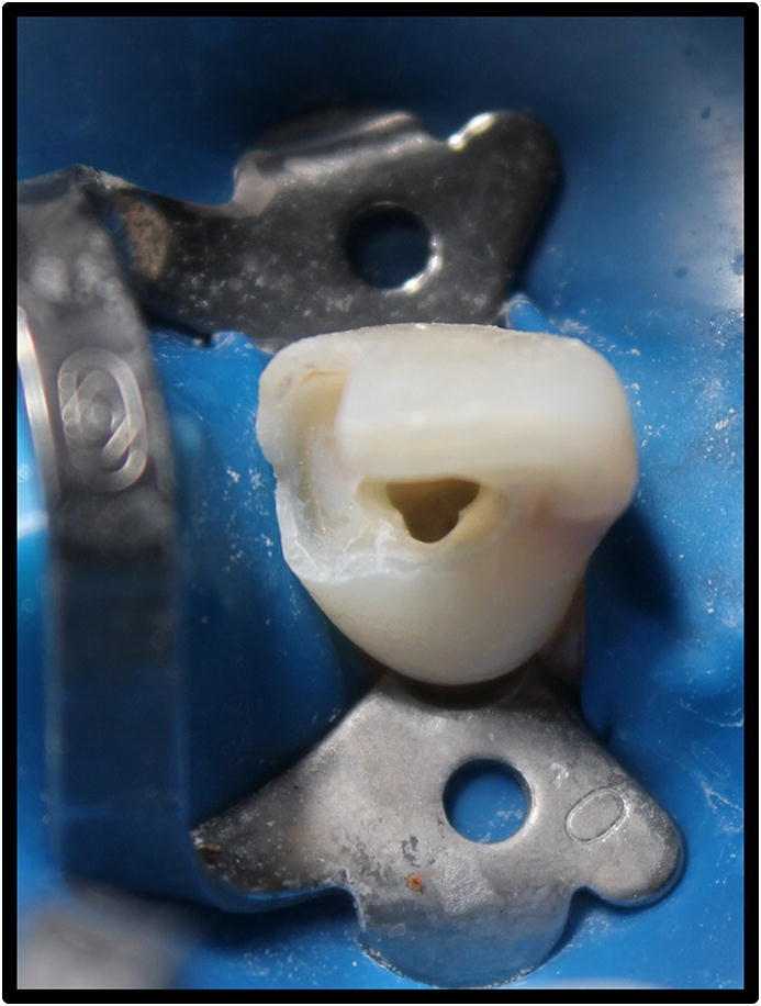
Rubber dam isolation and the appropriate access cavity.
Fig. 3.
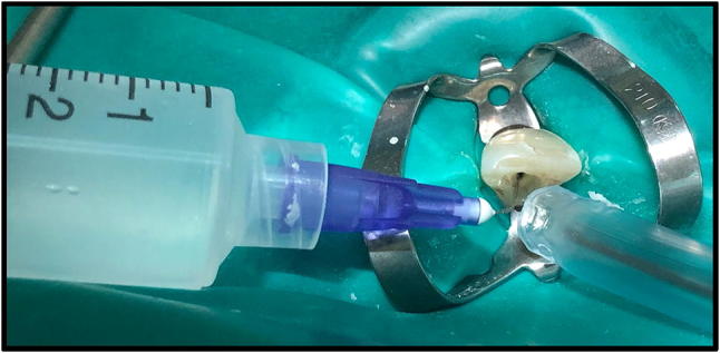
A final irrigation with 10 mL cold (1.5–2.5 °C) normal saline, using a needle with gauge size 30.
Fig. 4.
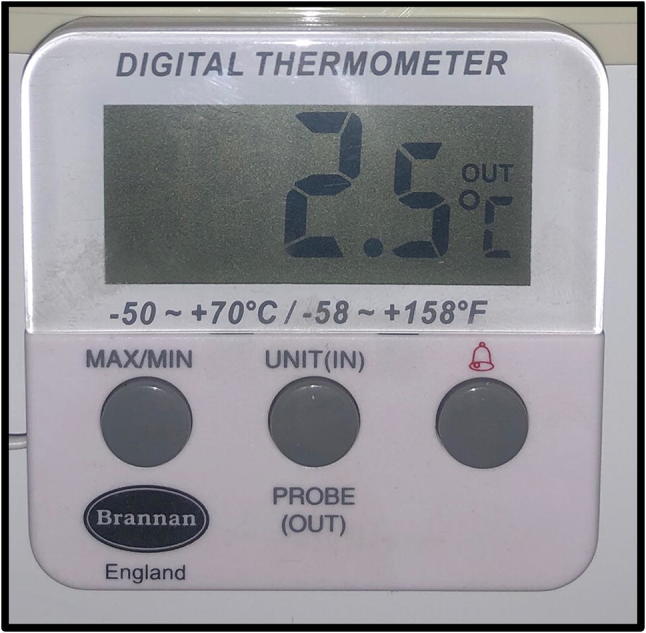
Digital thermometer to monitor the temperature of cold saline syringes inside the refrigerator.
Fig. 5.
Coronal access cavities restored with light cure Glass Ionomer restoration.
Fig. 6.
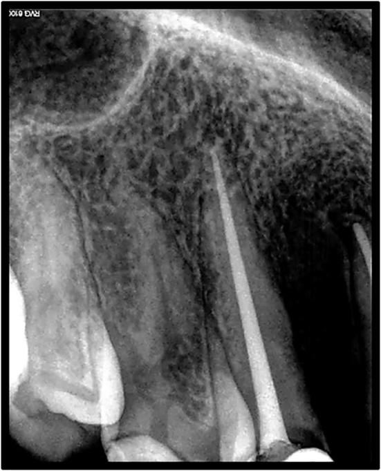
A periapical radiograph to ensure the obturation.
The patients were asked to fill out the VAS questionnaire and register their post-endodontic pain at 6, 24, and 48 h and instructed to contact their practitioner by phone to report whether they experienced post-endodontic pain. The patients who suffered from severe pain the investigators prescribed analgesic and the patient marked 10 on VAS.
Data were collected and analyzed by Statistical Package for the Social Sciences (SPSS) 13.0 using the Kolmogorov-Smirnov test to assess normal distribution. One-way analysis of variance (ANOVA) and was used to determine statistical difference (α = 0.05) among the groups. Tuckey’s test was used to calculate P-value (α = 0.05) between two each group.
3. Results
A total of 105 participants filled out the VAS questionnaire completely. Cryotherapy group (n = 35), room-temperature normal saline group (n = 35) and control group (n = 35).
The mean of post-endodontic pain level for all groups at 6, 24, and 48 h was calculated (Fig. 7).
Fig. 7.

A chart showing the mean of post-endodontic pain level results.
One-way analysis of variance (ANOVA) was used to determine statistical difference (α = 0.05) among the groups (Table 1).
Table 1.
P-value calculated with ANOVA test. P ≤ 0.005 is considered statistically significant.
| 6 h Post-Operative (Mean ± SD) | 24 h Post-Operative (Mean ± SD) | 48 h Post-Operative (Mean ± SD) | |
|---|---|---|---|
| Group 1 | 0.786 ± 1.369 | 0.429 ± 0.756 | 0.071 ± 0.267 |
| Group 2 | 1.143 ± 1.657 | 0.786 ± 1.369 | 0.143 ± 0.535 |
| Group 3 | 3.714 ± 2.984 | 3.000 ± 3.109 | 2.429 ± 2.936 |
| P-value | 0.006 | 0.001 | 0.001 |
Tuckey’s test was used to calculate P-value (α = 0.05) between two each group (Table 2).
The results showed that Group 1 had the lowest post-endodontic pain (6 h, 24 h, and 48 h). Nevertheless, there was no significant difference between Group 1 and Group 2.
The highest post-endodontic pain (6 h, 24 h, and 48 h) was in Group 3.
There was a significant difference in post-endodontic pain (6 h, 24 h, and 48 h) of Group 3 when compared with Group 1 and Group 2.
There was no significant difference in post-endodontic pain (6 h, 24 h, and 48 h) between vital and non-vital teeth in all groups.
In general, the post-endodontic pain (6 h, 24 h, and 48 h) decreased with time in all groups.
Table 2.
P-value calculated by Tuckey’s test. Multiple comparison between two each group.
| Multiple Comparison | 6 h Post-Operative | 24 h Post-Operative | 48 h Post-Operative |
|---|---|---|---|
| Group 1 Vs Group 2 | 0.871 | 0.966 | 0.989 |
| Group 1 Vs Group 3 | 0.006 | 0.002 | 0.002 |
| Group 2 Vs Group 3 | 0.016 | 0.001 | 0.002 |
4. Discussion
This randomized controlled study was conducted to evaluate the effect of irrigation solutions’ temperature on post-endodontic pain.
The limitations faced in this study were as follows: The number of participants who met the inclusion criteria were limited, the pain level was subjectively determined because no device for measure the pain. The most endodontic sealer can cause chemical irritation. So, the investigators chose Zinc Oxide Eugenol sealer.
The investigators excluded any cases with endodontic mishaps and sealer puff to ensure no external effects on post-endodontic pain, and they used Zinc Oxide Eugenol sealer; they did not use intra-canal medication to prevent chemical irritation (Siqueira and Barnett, 2004, AlRahabi, 2017).
Teeth with periapical pathosis have a higher risk to develop pain (Law et al., 2015), and symptomatic teeth have more chance to develop post-endodontic pain regardless of the pulp status (Tanalp et al., 2013). Cases with preoperative pain or periapical pathosis were excluded in this study because investigators excluded any other factors effect on post-endodontic pain and to standardize the cases. If these cases included, the cases with preoperative pain or periapical may give unreliable results.
Post-endodontic pain in this study decreased in all groups with time. These results are in accordance with Pak and White (2011), who found that the prevalence of post-endodontic pain is high but drops within 1 day after RCT and continues going down to reach a minimal level (Pak and White, 2011).
In this study, tooth vitality showed nonsignificant difference in post- endodontic pain. However, these findings were not discussed in this study. These results are not in accordance with findings by Sipavičiūtė and Manelienė (2014), who found that 47–60% of patients with asymptomatic necrotic pulp tissue have post-endodontic pain, although the pulp status effect on post-endodontic pain is controversial (Sipavičiūtė and Manelienė, 2014).
Post-endodontic pain control is important in root canal treatment (8). Ibuprofen is an effective analgesic to control post-endodontic pain according to recent systematic review and meta-analysis of NSAIDs for managing post- endodontic pain (Smith et al., 2017). The last irrigation with normal saline at either cold or room temperature in this study was an excellent choice for controlling post-endodontic pain and may be a good alternative to the use of analgesics. The normal saline in both Group 1 and Group 2 can flush and remove the remnants of NaOCl irrigation, because these remnants can cause chemical irritation and post-endodontic pain (Siqueira and Barnett, 2004), and the cleaning ability of normal saline might be an underlying source of post-endodontic pain reduction.
In the cryotherapy group, the investigators used saline at 2.5 °C because the root surface temperature can be reduced by more than 10 °C by using 2.5 °C saline irrigant for 5 min, according to in vitro study (Vera et al., 2015). The investigators used 10 mL of cold saline in the final irrigation, with gauge size 30 in the cryotherapy group to prolong the presence of the cold irrigant inside the canal for around 5 min. In Group 2, the investigators used 10 mL of saline at room temperature in the final irrigation with gauge size 30 to standardize the final irrigation protocol in both groups (Group 1 and Group 2) and made the difference in the irrigant temperature only.
According to two recent studies that conducted cryotherapy research of single-visit root canal treatment (RCT), pain level was reduced compared to the control group (Al-Nahlawi et al., 2016, Keskin et al., 2017). Vera et al. (2018) applied cryotherapy on 105 patients with necrotic pulp and found that post-endodontic pain can be minimized by using intra-canal cryotherapy. In this study, Intra-canal cryotherapy (Group 1) gave better results than room- temperature normal saline (as in Group 2) as the last irrigation, but statistically there was no significant difference between the two groups. These results are not in accordance with Al-Nahlawi et al., Keskin et al., and Vera et al., who found that cold normal saline was more effective on post-endodontic pain than room-temperature normal saline as the last irrigation (Al-Nahlawi et al., 2016, Keskin et al., 2017, Vera et al., 2018).
EndoVac was not used in this study because it is not available in all dental clinics, although Al-Nahlawi et al., Keskin et al., and Vera et al. used EndoVac for delivering the irrigation solution (Al-Nahlawi et al., 2016, Keskin et al., 2017, Vera et al., 2018).
Group 3, in the current study, had NaOCl irrigation without any additional saline in the regimen; it is regarded as the classic irrigation protocol (control). This yielded a statistically significant difference between Group 3 and Group 1 and Group 2 separately. Group 3 was not addressed in Al-Nahlawi et al., Keskin et al., and Vera et al., who compared only cold normal saline with room temperature normal saline as the last irrigation (Al-Nahlawi et al., 2016, Keskin et al., 2017, Vera et al., 2018).
Three participants in this study, every one of them had three teeth with single canal (Split-mouth study) which every tooth had been endodontically treated and allocated in different group (Group 1, Group 2 and Group 3). The teeth allocated in Group 1 and Group 2 were the least post-endodontic pain level and they did not feel difference between two methods. The teeth allocated in Group 3 were the highest post-endodontic pain level. This Split-mouth cases can reduce the inter-individual variability (Lesaffre et al, 2009).
5. Conclusion
Within the limitations of this study, we can conclude the following.
-
•
Final flushing of the canal with saline at either cold or room temperature was effective for post-endodontic pain control. This can be promising as an essential step in endodontic treatment to reduce the post-endodontic pain.
-
•
Room-temperature saline as the final irrigation showed results comparable to intracanal cryotherapy.
-
•
Further clinical studies should be done to determine the minimal volume of normal saline needed to control post-endodontic pain.
Acknowledgement
Authors would like to thank Dr. Ateeq Ayash Alotaibi, Dr.Ali Salem Hamdi, Dr. Bashar Aziz Alzubaidy, Duaa Salih Bazaid, Ghaida Raja Althebeti. And Dr. Wajdi Mohammed Bardisi for helping in collecting study sample patients.
Footnotes
Peer review under responsibility of King Saud University.
References
- Al-Nahlawi T., Hatab T.A., Alrazak M.A., Al-Abdullah A. Effect of intracanal cryotherapy and negative irrigation technique on postendodontic pain. J. Contemp. Dent. Pract. 2016;17(12):990–996. [PubMed] [Google Scholar]
- AlRahabi M. Predictors, prevention and management of postoperative pain associated with nonsurgical root canal treatment: a systematic review. J. Taibah. Univ. Med. Sc. 2017;12(5):376–384. doi: 10.1016/j.jtumed.2017.03.004. [DOI] [PMC free article] [PubMed] [Google Scholar]
- Keskin C., Ozdemir O., Uzun I., Guler B. Effect of intracanal cryotherapy on pain after single-visit root canal treatment. Aust. Endod. J. 2017;43(2):83–88. doi: 10.1111/aej.12175. [DOI] [PubMed] [Google Scholar]
- Law A., Nixdorf D., Aguirre A., Reams G., Tortomasi A., Manne B. Dental PBRN collaborative group; predicting severe pain after root canal therapy in the national dental PBRN. J. Dent. Res. 2015;94(3 Suppl):37S–43S. doi: 10.1177/0022034514555144. [DOI] [PMC free article] [PubMed] [Google Scholar]
- Lesaffre E., Philstrom B., Needleman I., Worthington H. The design and analysis of split-mouth studies: what statisticians and clinicians should know. Statist. Med. 2009;28:3470–3482. doi: 10.1002/sim.3634. [DOI] [PubMed] [Google Scholar]
- Mandras N., Allizond V., Banche G., Roana J., Piazza L., Viale P., Cuffini A.M. Antimicrobial efficacy of cryotreatment against enterococcus faecalis in root canals. Lett. Appl. Microbiol. 2013;56(2):95–98. doi: 10.1111/lam.12017. [DOI] [PubMed] [Google Scholar]
- Manfredi M., Figini L., Gagliani M., Lodi G. Single versus multiple visits for endodontic treatment of permanent teeth. Cochrane Database Syst. Rev. 2016;12 doi: 10.1002/14651858.CD005296.pub3. [DOI] [PMC free article] [PubMed] [Google Scholar]
- Nadler S., Weingand K., Kruse R. The physiologic basis and clinical applications of cryotherapy and thermotherapy for the pain practitioner. Pain. Physician. 2004;7:395–399. [PubMed] [Google Scholar]
- Pak J., White S. Pain prevalence and severity before, during, and after root canal treatment: a systematic review. J. Endod. 2011;37(4):429–438. doi: 10.1016/j.joen.2010.12.016. [DOI] [PubMed] [Google Scholar]
- Sipavičiūtė E., Manelienė R. Pain and flare-up after endodontic treatment procedures. Baltic Dental Maxillofacial J. 2014;16:25–30. [PubMed] [Google Scholar]
- Siqueira J., Barnett F. Interappointment pain: mechanisms, diagnosis and treatment. Endod. Topics. 2004;7:93–109. [Google Scholar]
- Smith E., Marshall J., Selph S., Barker D., Sedgley C. Nonsteroidal anti-inflammatory drugs for managing postoperative endodontic pain in patients who present with preoperative pain: a systematic review and meta-analysis. J. Endod. 2017;43:7–15. doi: 10.1016/j.joen.2016.09.010. [DOI] [PubMed] [Google Scholar]
- Tanalp J., Sunay H., Bayirli G. Cross-sectional evaluation of post-operative pain and flare-ups in endodontic treatments using a type of rotary instruments. Acta Odontol dScand. 2013;71:3–4. doi: 10.3109/00016357.2012.715199. [DOI] [PubMed] [Google Scholar]
- Vera J., Ochoa J., Romero M., Vazquez-Carcan O.M., Ramos-Gregorio C., Aguilar R., Cruz A., Sleiman P., Arias A. Intracanal cryotherapy reduces postoperative pain in teeth with symptomatic apical periodontitis: a randomized multicenter clinical trial. J. Endod. 2018;44:4–8. doi: 10.1016/j.joen.2017.08.038. [DOI] [PubMed] [Google Scholar]
- Vera J., Ochoa J., Vazquez-Carcaño M., Romero M., Arias A., Sleiman P. Effect of intracanal cryotherapy on reducing root surface temperature. J. Endod. 2015;41(11):1884–1887. doi: 10.1016/j.joen.2015.08.009. [DOI] [PubMed] [Google Scholar]