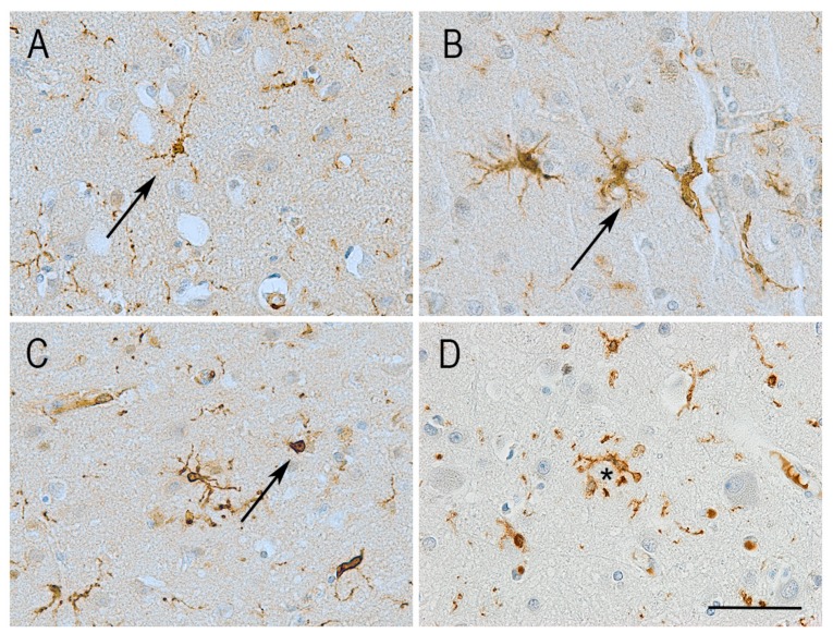
Figure 1.
Illustration of the different morphologies adopted by microglia in the human brain independently of age or disease. Immunolabelling for the microglial protein Iba1 shows diverse morphologies including varying number of processes and cell body shape. (A) Ramified microglia with small round cell body and several long branching processes. (B) Reactive microglia with increased cell body size and reduced length of processes. (C) Amoeboid microglia with enlarged cell body and no processes. Images A–C taken from a control aged brain. (D) Microglia clustering around Aβ plaques (*) is a feature observed only in the presence of Alzheimer-type pathology. Haematoxylin counterstaining. Scale bar = 50 μm.

