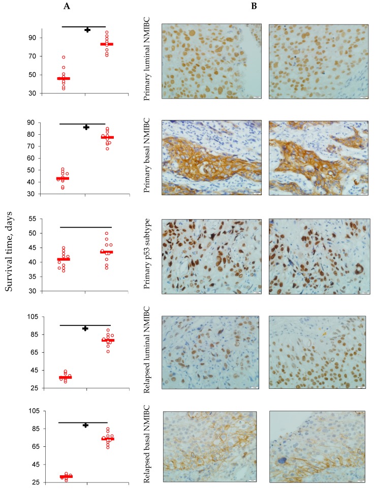
Figure 2.
(A) Average survival of animals in experimental groups; average survival (n = 10) presented in days of life (scatterplots and median); ÷ p < 0.05 when compared with control (Gehan’s criterion with Yates’s correction). (B) Expression of GATA 3 (1, 4, 7, and 10), KRT 5/6 (2, 5, 8, and 11), and p53 (3, 6, 9, and 12) in maternal tumor’s specimens and in established PDX’s ones; IHC staining, ×600.


