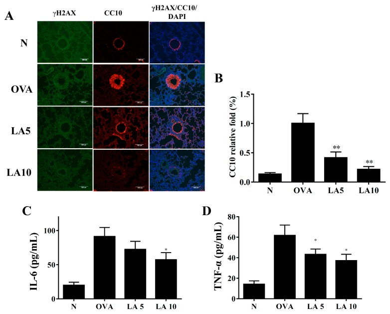
Figure 7.
Licochalcone A (LA) affected DNA damage in asthmatic lung tissue. (A) Histological sections of lung tissues were stained for γH2AX (green) and CC10 (red) by immunofluorescence. Nuclei were stained with DAPI (blue) and observed by fluorescence microscopy. (B) Fold-changes in the expression levels of CC10 were measured and compared to the OVA group. Effects of LA on the levels of proinflammatory cytokines in BALF. (C) The concentrations of IL-6 and (D) TNF-α in BALF were measured by ELISA. Three independent experiments were analyzed, and data were presented as mean ± SEM. * P < 0.05 compared to the OVA control group. ** P < 0.01 compared to the OVA control group.

