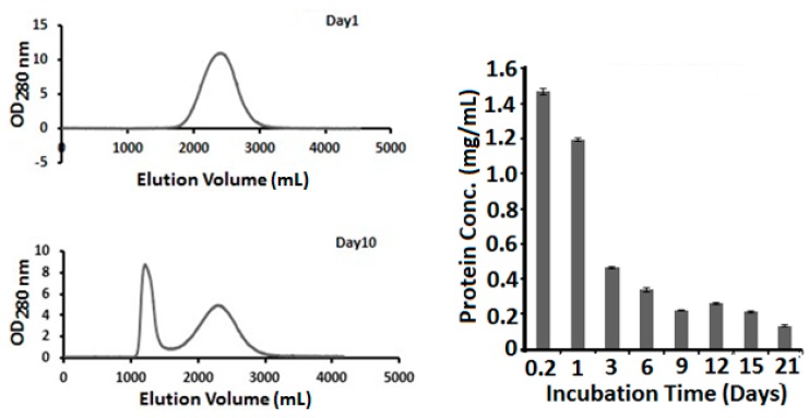
Figure 2.
Non-fibrous lysozyme. As fibers and gel are formed, a substantial amount of lysozyme remains in non-fibrous form. (Left Panel) fast protein liquid chromatograpy (FPLC) analysis of soluble non-fibrous lysozyme. After centrifugation to remove fibers from the amyloid fiber solution, supernatant was analyzed by FPLC. The peak eluted at around 1200 mL from the column is thought to be the colloidal peak, consisting mostly spheres and very short fibers. The peak eluated at around 2300 mL is thought to be lysozyme monomers. (Right Panel) Non-fibrous lysozyme concentrations were measured for lysozyme fiber reactions harvested at different times.

