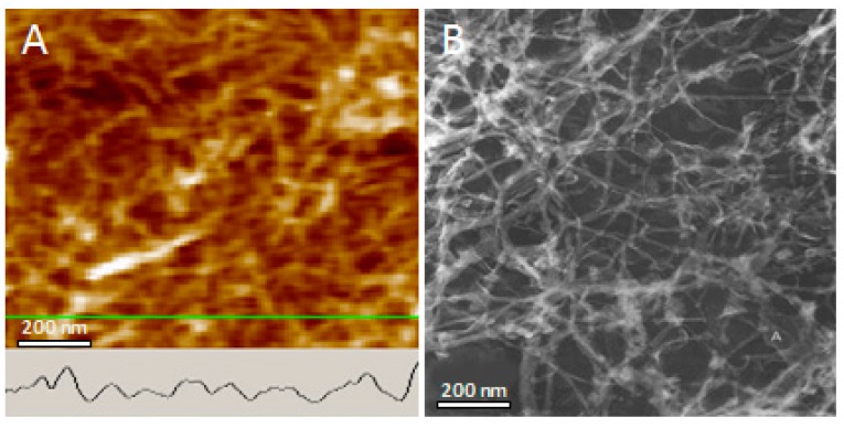
Figure 5.
Network formed by lysozyme amyloid fibers (AFM, topographic view) and by tau neurofibrillary tangles (TEM image from Ruben, 1997). The AFM profile of lysozyme fiber is shown (inset). Distance between fibers and pore size is similar in both. Merging of fibers at points of contact suggest coiling of fibers with each other in both cases.

