Figure 2.
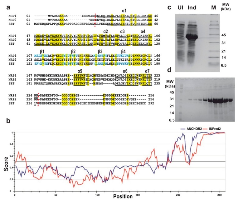
Expression, purification, and sequence analysis for AtNRP2. (a) Sequence alignment of AtNRP2 with AtNRP1 and HsSET/TAF-1β. All the conserved residues are shown in yellow. Secondary structures of known structures are shown; wherein, α-helices are underlined, and β-sheets are in blue and italics. Red dotted lines mark the regions where AtNRP2 was truncated based on the secondary structure information of the other two proteins. (b) IUPRED prediction of disordered regions of AtNRP2. (c) SDS-PAGE imageshowing the expression of AtNRP2 NT/CT upon IPTG induction. The lanes are marked as UI = uninduced, Ind = Induced, and M = Marker. (d) SDS-PAGE image showing the purified peak fractions of AtNRP2 NT/CT from gel-filtration chromatography using HiLoad 16/600 Superdex 200pg column (GE Healthcare).The extreme left lane has the marker, and the remaining lanes have the fractions of the protein from gel-filtration.

