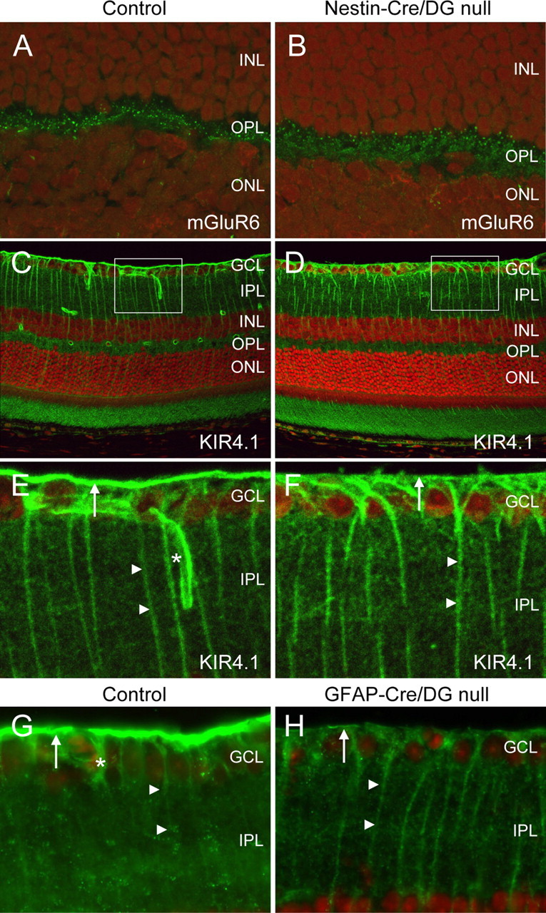
In the article “Visual Impairment in the Absence of Dystroglycan” by Jakob S. Satz, Alisdair R. Philip, Huy Nguyen, Hajime Kusano, Jane Lee, Rolf Turk, Megan J. Riker, Jasmine Hernández, Robert M. Weiss, Michael G. Anderson, Robert F. Mullins, Steven A. Moore, Edwin M. Stone, and Kevin P. Campbell, which appeared on pages 13136–13146 of the October 21, 2009 issue, there was an error in Figure 7. The labels for the Outer Plexiform Layer (OPL) were mislabeled as Ganglion Cell Layer (GCL) in A and B. The correct version of Figure 7 appears below.
Figure 7.

Partial limbic seizures in pilocarpine-treated rats are associated with fast polyspike activity in the hippocampus and ictal neocortical slow activity in the frontal cortex. Spontaneous seizures are shown from chronic video/EEG recordings in rats with pilocarpine-induced epilepsy. A, LFPs in the hippocampus and orbitofrontal cortex (CTX) during a Racine class 0 partial seizure of 50 s length, associated with behavioral arrest but no convulsive activity. Hippocampal recordings reveal large-amplitude, fast polyspike activity during the seizure, whereas frontal recordings show large-amplitude 1–2 Hz slow waves during and after the seizure without considerable propagation of fast spike activity. Ictal and postictal slow activity resembles large-amplitude slow rhythms seen during an episode of natural sleep, recorded in the same animal at a different time (bottom, right). B, Hippocampal and frontal recordings during a 46 s Racine class 5 secondarily generalized seizure, associated with bilateral tonic-clonic convulsions and loss of balance. LFP recordings reveal large-amplitude, fast polyspike activity in both the hippocampus and frontal cortex during the seizure, without frontal slow oscillations. LFP recordings are filtered 0.3–100 Hz.


