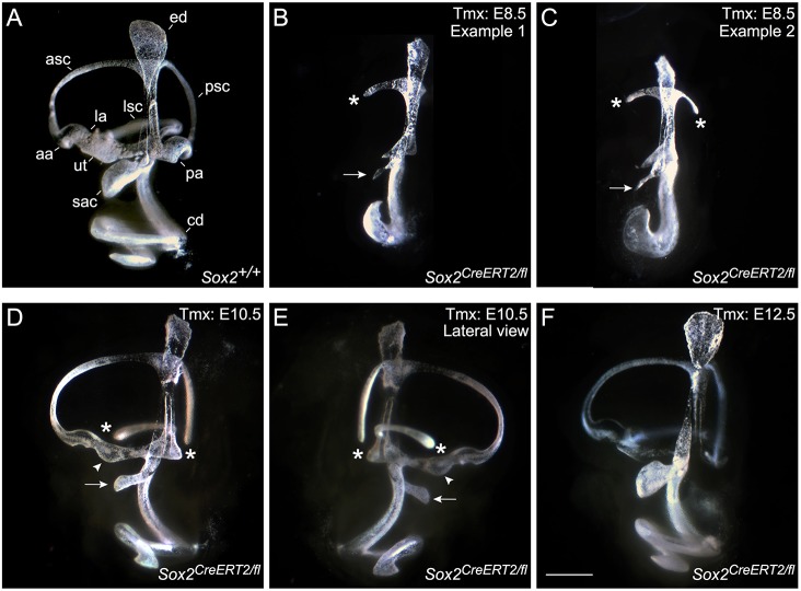
Fig. 3.
Paint-filling reveals a severe inner ear malformation resulting from the early deletion of SOX2, whereas later deletion has little effect on the overall morphology. (A-F) Paint-filled E15.5 inner ears with SOX2 deletion at indicated time points during development. Control (A) and two examples of E8.5 Sox2-deleted mutants (B,C) demonstrate severe loss of otic tissue caused by early loss of SOX2. (D,E) Medial and lateral views of an E10.5-deleted mutant show loss of the lateral and posterior ampullae, smaller maculae and an undercoiled cochleae. (F) Deletion of SOX2 at E12.5 did not show overt morphological defects. The smaller saccule (sac) is marked by an arrow (B-E), the utricle (ut) is marked by an arrowhead (D,E), missing ampullae or truncated canals are indicated by an asterisk (B-E). aa, anterior ampulla; asc, anterior semicircular canal; cd, cochlear duct; ed, endolymphatic duct; la, lateral ampulla; lsc, lateral semicircular canal; pa, posterior ampulla; psc, posterior semicircular canal. Scale bar: 500 μm.

