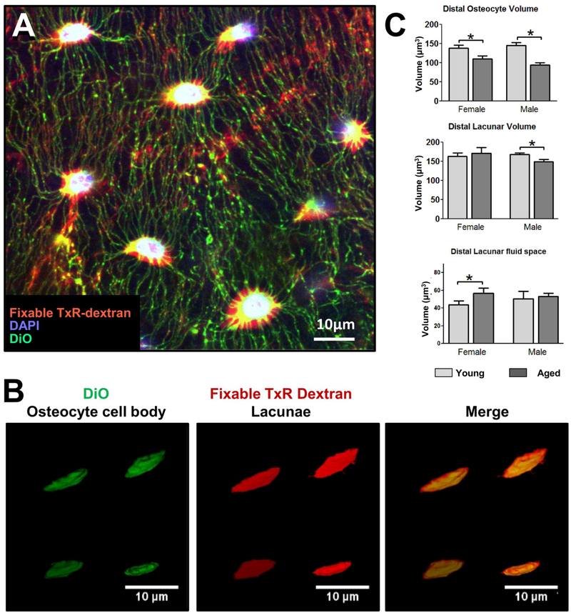
Figure 4: Multiplexed Imaging and Volumetric Quantitation of Osteocyte Cell body and Lacunae in Young and Aged Mice.
A) Multiplexed confocal imaging of the osteocyte cell body and lacunae in mouse femur using combined staining with fixable Texas Red-Dextran [red] to show the lacunocanalicular network, DAPI [blue], to show the nuclei and DiO [green], to show the cell membrane (single Z plane, 40x oil objective, 4x digital zoom), bar = 10μm. B) Examples of 3D renderings of osteocyte cell bodies labeled with DiO and lacunae labeled with fixable Tx-Red-Dextran for volumetric calculations, with merged image shown on the right, bar = 10μm. C) quantitation of osteocyte cell volume in the distal region of the femur in young and aged mice with quantitation of the corresponding lacunar volume and lacunar fluid space volume (Data are mean ± SEM, * = p< 0.05, ANOVA/Tukey’s; n= 5). Panels B) and C) are reproduced from Tiede-Lewis et al, 2017 [48].

