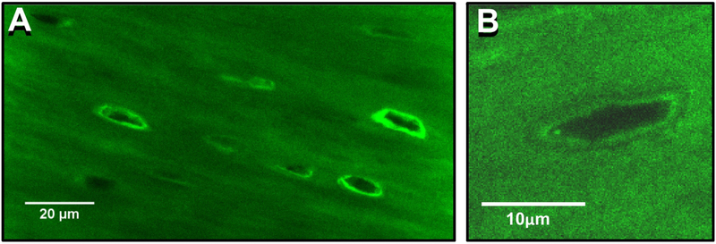
Figure 6: Evidence of Osteocyte Perilacunar Matrix Formation.
A) Confocal image from femoral cortical bone from a transgenic mouse expressing a GFP-tagged collagen construct. Note that there are numerous osteocyte lacunae surrounded by bright rings of GFP-collagen fluorescence, suggesting that the osteocytes may be able to add collagen to their perilacunar matrix, bar = 20μm (single Z-plane, 100x oil objective), bar = 20μm. B) Enlarged image of a single osteocyte lacuna from a GFP-collagen mouse showing interfaces around the lacuna that suggest GFP-collagen addition and removal, bar = 10μm.

