. 2019 Jul 8;8:e46156. doi: 10.7554/eLife.46156

Figure 2. Reduction in GM volume, rsALFF and rsFC in autoimmune LE patients.

(a) A whole-brain VBM on GM volume (contrast: controls > patients) showed volume reduction in patients’ HPC bilaterally, as well as in mediodorsal-anterior and right dorsolateral thalamic regions (Table 4); clusters survive FWE peak-level correction (p<0.05) over p<0.001 unc; color bar indicates t values; b-c: Reduced rsALFF in patients in (b) the posterior cingulate (kE = 89, p-FWE = 0.033; peak voxel coordinates: −4,–36, 28) and (c) precuneus (kE = 137; p-FWE = 0.003; peak voxel coordinates: 4,–60, 26); d-j: reduced rsFC in patients; d: a whole-brain MVPA (omnibus F) showed abnormal rsFC for patients in a cluster in the right HPC (kE = 178, p-FWE = 0.001; peak voxel coordinates: 28,–16,−20; color bar indicates F values); e-g: reduced rsFC of the right HPC (whole-brain seed-to-voxel analysis; seed: right HPC, anatomically delineated in native space, unsmoothed timeseries; contrast: controls > patients); e: medial prefrontal cortex (kE = 1152, p-FWE <0.0001, peak voxel coordinates: 4, 56, 2); f: posteromedial cortex (posterior cingulate, retrosplenial cortex, precuneus; kE = 986, p-FWE <0.0001, peak voxel coordinates: 6,–50, 8); g: left HPC (kE = 393, p-FWE <0.0001, peak voxel coordinates: −12,–36, 2). All rsFC and rsALFF clusters survive FWE correction (p<0.05) for cluster size over an individual voxel threshold of p<0.001; FWE: family-wise error; HPC: hippocampus; kE: cluster size (number of voxels); rsALFF: Resting-state amplitude of low frequency fluctuations; rsFC: Resting-state functional connectivity; VBM: voxel-based morphometry.

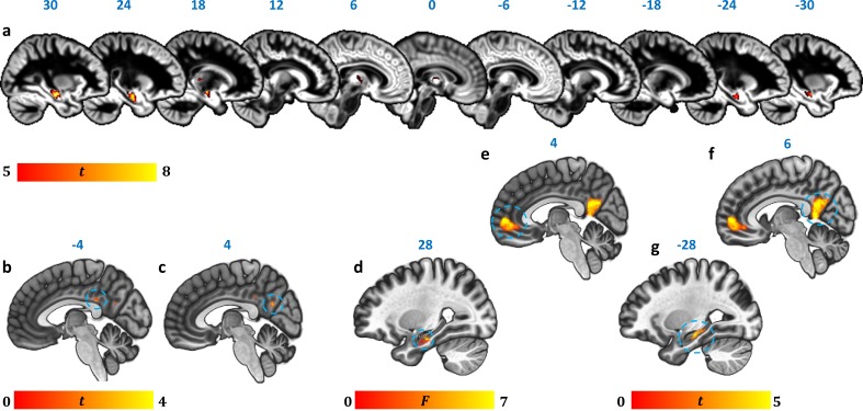
Figure 2.

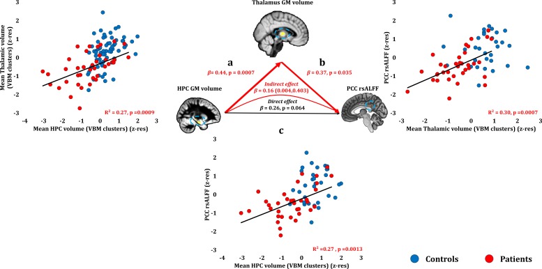
Figure 2—figure supplement 1. Relationship of HPC atrophy with PCC functional abnormalities.

Figure 2—figure supplement 1.

Given the reciprocal connectivity of thalamic nuclei with both the HPC and the cingulate cortex (Aggleton, 2014; Aggleton et al., 2010; Bubb et al., 2017) and our hypothesis that HPC atrophy is followed by structural and functional abnormalities in interconnected areas within the HPC-diencephalic-cingulate networks, we assumed a causal chain of events, whereby HPC damage has remote effects on thalamic volume, which, in turn, lead to abnormalities in the cingulate cortex, observed here in the form of reduced rsALFF in the PCC. We used a series of bivariate correlations and mediation analyses, with HPC volume as the independent variable, thalamic volume as the mediator variable, and rsALFF in the PCC as the dependent variable. A mediation analysis supported this hypothesis, showing that the effects of the average GM volume reduction in the HPC VBM clusters on patients’ reduced rsALFF in the PCC were fully mediated by the correlative reduction of the average GM volume of the thalamic VBM clusters (direct effect: β = 0.26, p=0.064; indirect effect: β = 0.16, 95% CI: 0.004,0.403); a: mean GM volume of the two HPC clusters correlated with the mean GM volume of the two thalamic clusters across patients; b: mean PCC rsALFF correlated across patients with the mean GM volume of the two thalamic clusters; the mediation analysis demonstrates that this effect held when the correlation of thalamic GM volume with the mean GM volume of the HPC clusters was accounted for; c: mean GM volume of the HPC clusters correlated with PCC rsALFF; the mediation analysis demonstrated that this relationship did not hold over and above the correlation of the mean GM volume of the thalamic clusters with the HPC clusters; there was thus no direct effect of reduced HPC GM volume on PCC rsALFF (within parenthesis: 95% confidence intervals); GM: gray matter; HPC: hippocampus; MAP: Memory and Amnesia Project; OPTIMA: Oxford Project To Investigate Memory and Aging; PCC: posterior cingulate cortex; rsALFF: resting-state amplitude of low frequency fluctuations; TIV: total intracranial volume; VBM: voxel-based morphometry; z-res: GM volumes from VBM clusters are residualized against age, sex, scan source (MAP, OPTIMA), and TIV across participants; mean rsALFF is residualized against age and sex across participants.
Figure 2—figure supplement 1—source data 1. This spreadsheet contains the mean GM volume of the HPC and thalamic VBM clusters and the mean rsALFF in the PCC cluster (z-res) for healthy controls and patients that are plotted in Figure 2—figure supplement 1; GM: gray matter; HPC: hippocampus; MAP: Memory and Amnesia Project; OPTIMA: Oxford Project To Investigate Memory and Aging; PCC: posterior cingulate cortex; rsALFF: resting-state amplitude of low frequency fluctuations; TIV: total intracranial volume; VBM: voxel-based morphometry; z-res: GM volumes from VBM clusters are residualized against age, sex, scan source (MAP, OPTIMA), and TIV across participants; mean rsALFF is residualized against age and sex across participants.
These data can be opened with Microsoft Excel or with open-source alternatives such as OpenOffice.
DOI: 10.7554/eLife.46156.007