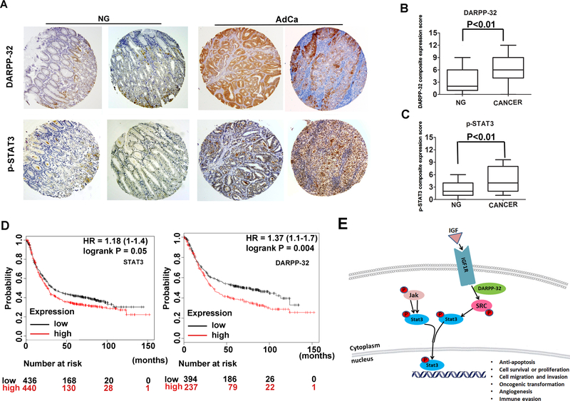
Figure 7. Immunohistochemistry for DARPP-32 and p-STAT3 in human gastric tissues.
A) Representative images of immune-histochemical staining of DARPP-32 and p-STAT3 in tissue sections from human gastric mucosa with normal histology (NG, n=108) and adenocarcinoma (AdCa, n=108); original magnification ×20. B-C) The graphs summarize the immunohistochemical staining results on gastric tissue microarrays. D) Survival analysis of DARPP-32 and STAT3 mRNA expression in gastric cancer patients by the Kaplan-Meier survival curve, n=882, following analysis of public data online (http://kmplot.com/analysis/index.php?p=service) [25]. E) A diagram depicting the role of DARPP-32 in activation of STAT3 in gastric cancer cells. In summary, DARPP-32 interacts with IGF1R and promotes IGF1R and SRC phosphorylation, allowing sustained IL6-mediated phosphorylation and activation of STAT3. The translocation of p-STAT3 into the nucleus initiates transcriptional regulation of downstream target genes that regulate cell proliferation, transformation.

