Abstract
Purpose
Intraocular pressure leading to glaucoma is a major cause of childhood blindness in developing countries. In this study, we sought to identify gene variants potentially associated with primary congenital glaucoma (PCG) in the Mauritanian population.
Methods
Using next-generation sequencing (NGS), a panel of PCG candidate genes was screened in a search for DNA mutations in four families with multiple occurrences of PCG.
Results
Targeted exome sequencing analysis revealed predicted pathogenic mutations in four genes: CYP1B1 (c.217_218delTC, p.Ser73Valfs*150), MYOC (878C>A, p.T293K), NTF4 (c.601T>G, p.Cys201Gly), and WDR36 (c.2078A>G, p.Asn693Ser), each carried by a different family.
Conclusions
Genetic variation associated with PCG in this study reflects the ethnic heterogeneity of the Mauritanian population. However, a larger cohort is needed to identify additional families carrying these mutations and confirm their biologic role.
Introduction
Glaucoma mainly results from a slow, asymptomatic, and bilateral build-up of pressure in the eye, as the fluid drainage channels become progressively blocked, leading to damage of the optic nerve [1]. In less frequent forms, the presence of increased intraocular pressure is congenital, occurring in about 1 out of 10,000 births in Western populations [2]. Glaucoma manifests during the neonatal or early infantile period [3]. In developing countries, as the disease often goes undetected or untreated in the childhood, damage to the optic nerve often leads to irreversible corneal opacification, and eventually, permanent visual loss.
Most cases of primary congenital glaucoma (PCG) are sporadic. However, its prevalence among consanguineous families and some ethnic groups supports a genetic basis for this disorder [4]. The inheritance is predominantly autosomal recessive, although pedigrees with a dominant transmission mode have also been described [5]. Mutations in different genes, largely CYP1B1 (GLC3A, 2p22-p21; Gene ID:1545; OMIM 601771), but also MYOC (Gene ID:4653; OMIM 601652), LTBP2 (Gene ID:4053; OMIM 602091), PITX2 (Gene ID:5308; OMIM 601542), FOXC1 (Gene ID:2296; OMIM 601090), and PAX6 (Gene ID:5080; OMIM 607108), have been associated with congenital glaucoma cases in various populations [6-9]. However, data on underlying genetic variants in African populations remain limited [10]. As part of a comprehensive study aimed at investigating the genetic etiology of vision impairment in the Mauritanian population [11], this study examined, for the first time, the mutation spectrum of the main glaucoma candidate genes in Mauritanian families with PCG.
Methods
Selection of families
Initially, 16 visually impaired children, from 16 unrelated families, attending a school for the blind in Nouakchott, the capital city, were recruited. Age at onset of the disease and family medical history were recorded following interviews with the parents. Ophthalmological examination of the patients included visual acuity measurement, slit lamp, tonometry, and fundoscopy. Pedigrees of the four families with more than one patient with PCG were charted. Other ocular abnormalities, such as cataract, were excluded.
Targeted exome sequencing
DNA extraction from peripheral blood collected in EDTA tubes was performed using the QIAamp DNA Blood Mini Kit (Qiagen, Hilden, Germany). Venous blood was drawn in EDTA- containing vacutainer tubes and used for DNA extraction within hours from the time of collection. For longer term storage, samples were kept at -20 °C. Genomic DNA was extracted using the QIAamp DNA Blood Mini Kit (Qiagen, Hilden, Germany) following manufacturer’s instructions. Underlying mutations were first searched in the probands with next-generation sequencing (NGS) using a custom-designed in-solution capture array targeting a panel of 55 glaucoma candidate genes. Details of the relevant genes are provided in Appendix 1. Library preparation, qualification, and NGS on the HiSeq 2000 platform (Illumina, Inc., San Diego, CA) were performed as described previously [10]. Briefly, after DNA extraction, shearing, ligation, library selection and hybridization, targeted captured multiplex DNA samples were then amplified by ligation-mediated PCR (LM-PCR) using SeqCap EZ Accessory Kit v2 (Roche, Swiss) with the following program: 98 °C×45 s, (98 °C × 15 s, 60 °C × 30 s, 72 °C×30 s) × 13 cycles, and hold at 4 °C after 72 °C×1 min. After purification, reads sequencing on amplified captured multiplex DNA sample was then performed followed by assembly of the sequences and gene annotation.
Direct Sanger sequencing was subsequently performed to confirm the NGS data and familial segregation. Individuals used as controls included sighted persons from the four enrolled families and other PCG-unaffected subjects from the general population. In total, DNA from 15 controls (visually non-impaired subjects) was analyzed using the same sequencing approach. Details of the designed relevant PCR primers are shown in Table 1.
Table 1. List of genes and primer sequences used to detect mutations in PCG patient DNAs.
| Gene | Location | Primers |
|---|---|---|
| CYP1B1 | Exon 2 | F:TCGCCATTCAGCACCACTAT |
| |
|
R:CTCCACCCAACGGCACTCAG |
| MYOC | Exon 3 | F:TGGCTACACGGACATTGACT |
| |
|
R:TAGCTGCTGACGGTGTACAA |
| NTF4 | Exon 2 | F:CCAAAACCCCATGTGGTTTC |
| |
|
R:GCTGTGTGCGATGCAGTCAG |
| WDR36 | Exon 18 | F:ACACAGCTTTGTTATGATGGCA |
| R:GCCTTTGGTAGCAATATCTCCT |
Bioinformatics analyses
In the reads mapping against the human genome 19 (hg19) reference sequence to reveal potential genetic differences, we used single nucleotide polymorphism (SNP) databases such as HapMap project, 1000 Genome Project, Exome Variant Server (EVS), Exome Aggregation Consortium (EXAC), and Genome Aggregation Database (GnomAD). Alamut Visual 2.11 (Interactive Biosoftware, Rouen, France) was used to estimate the pathogenicity of detected variants. The amino acid change was considered potentially disease causing if predicted by at least one of the programs (PolyPhen2), Sorting Intolerant From Tolerant (SIFT), and Mutation Taster. Amino acid conservation across species was studied with the UCSC Genome Browser.
Approval for this study was given by the ethics committee of the University of Sciences, Technologies and Medicine, Nouakchott, Mauritania. The purpose of the study was explained to the participants, and their informed and signed consent taken. For children, the parents’ approval was obtained. This study was carried out in accordance with the ethical principles for medical research involving human subjects defined by the World Medical Association Declaration of Helsinki.
Results
Clinical exploration
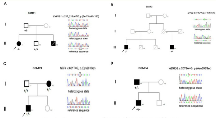
Out of the 16 visually impaired children, from 16 recruited families, bilateral PCG was confirmed or newly diagnosed in only four families, as affected children from these families were found to have at least one other PCG patient relative. They were subsequently termed blindness by glaucoma in Mauritanian families (BGMFs). In all patients, signs of vision loss were noticed in the first year of life. They presented photophobia, epiphora, corneal clouding, and enlargement of the globe or cornea. The extent of impairment varied initially but led to total blindness in all PCG cases. Pedigrees (Figure 1) were consistent in two families (BGMF1 and BGMF2) with autosomal recessive inheritance. In the two other families, an autosomal dominant pattern was observed. No signs of additional ocular deficiency or other organ disease were diagnosed in the patients.
Figure 1.

Family pedigrees and DNA partial sequences showing mutations in respective genes. A: Deletion (c.217_218delTC) in exon 2 of CYP1B1 (OMIM: 601771) in blindness by glaucoma in Mauritanian family 1 (BGMF1). B: Missense mutation (c.878C>A) in exon 3 (rs139122673) of MYOC (OMIM: 60165) in BGMF2. C: Missense mutation (c.601T>G) in exon 2 of NTF4 (OMIM: 162662) in BGMF3. D: Missense mutation (c.2078A>G) in exon 18 of WDR36 in BGMF4.
Genetic screening
Mutation analysis of data from targeted exome sequencing performed on the DNA of the probands from the four selected families showed sequence variations in different genes. Each family affected with PCG carries only one mutant gene (Table 2). In BGMF1, a homozygous mutation in CYP1B1 (OMIM: 601771) was detected in the proband (a 10-year-old girl). This 2 bp deletion (c.217_218delTC) occurred in exon 2 and created a frame shift at the conserved Ser73 codon. The new reading frame brought on a terminating codon at position 150 downstream (p.Ser73Valfs*150). The homozygous mutation was confirmed in the proband, and identified in her brother, also blind due to PCG, with Sanger sequencing. This mutation was also detected, in the heterozygous state, in the family’s father who was visually unimpaired (Figure 1A).
Table 2. List of variations detected by NGS in Mauritanian PCG families.
| ID | Ethnic origin | Gene | Mode of inheritance | Location | Nucleotide change | Allelic form | Amino acid change | Frequencies (ESP, ExAc, GnomAd) | Polyphen2 | SIFT | Reference |
|---|---|---|---|---|---|---|---|---|---|---|---|
| BGMF1 |
white Maure |
CYP1B1 |
AR |
Exon 2 |
c.217_218del |
Homo
zygous |
p.(Ser73Valfs*150) |
ExAc:AALL:0.00057%-AFR:0.010%-AMR:0%-EAS:0%-SAS:0%-NFE:0%-FIN:0%-OTH:0%; |
n.a. |
n.a. |
Chen et al., 2014 |
| BGMF2 |
white Maure |
MYOC |
AR |
Exon 3 |
c.878C>A |
Homo
zygous |
p.(Thr293Lys) |
ExAc: ALL:A=0.058%-AFR:0.020%-AMR:0.13%-EAS:0%-SAS:0%-NFE:0.057%-FIN:0%-OTH:0.13%; ESP: EA: T=0.05% - AA: T=0.07%; GnomAd: 0.0005603, never homozygous |
Benign |
Tolerated |
rs139122673 |
| BGMF3 |
black African |
NTF4 |
AD |
Exon 2 |
c.601T>G |
Hetero
zygous |
p.Cys201Gly |
- |
Probably damaging |
Deleterious |
Novel |
| BGMF4 | black African | WDR36 | AD | Exon 18 | c.2078A>G | Hetero zygous | p.Asn693Ser | ALL:G=0.0024%-AFR:0.020%-AMR:0.0089%-EAS:0%-SAS:0%-NFE:0%-FIN:0%-OTH:0%; GnomAD: 0.00002394 never homozygous | Probably damaging | Deleterious | rs752189803 |
n.a. not applicable 1- ZhaoY,SorensonCM,SheibaniN.CytochromeP4501B1and 2- primary congenital glaucoma. J Ophthalmic Vis Res 2015; 10: 3- 60–6 4- ZhaoY,SorensonCM,SheibaniN.CytochromeP4501B1and 5- primary congenital glaucoma. J Ophthalmic Vis Res 2015; 10: 6- 60–6
A second homozygous mutation, in the MYOC gene (OMIM: 601652), was detected in the proband of the BGMF2 family. This missense mutation (c.878C>A) in exon 3 (rs139122673) was also identified in his cousin, with Sanger sequencing. The mutation provoked the replacement of threonine by a lysine amino acid (p.T293K; Figure 1B). Both mutations (c.217_218delTC) in CYP1B1 and (c.878C>A) in MYOC were classified as rare variants based on their respective minor allele frequency (MAF) in the ExAc database (Table 2).
A heterozygous missense mutation (c.601T>G) in exon 2 of NTF4 (OMIM: 162662) translating at the protein level in p.Cys201Gly was also found and confirmed with Sanger sequencing in two blind members of BGMF3. The proband and his mother were both affected by bilateral PCG (Figure 1C).
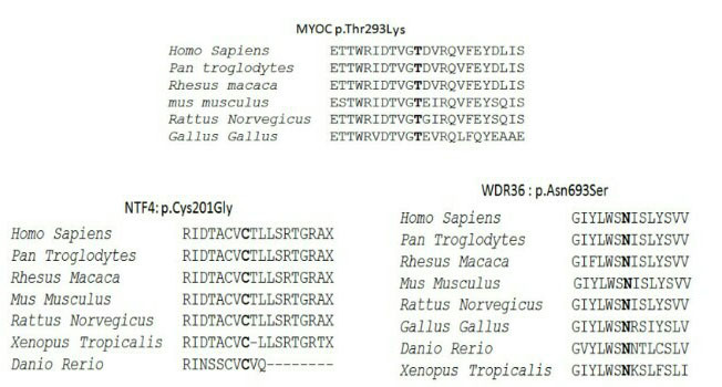
In BGMF4, another heterozygous mutation in exon 18 of WDR36 (c.2078A>G, p.Asn693Ser) was carried by a 13-year-old boy and his father; both were patients with PCG. This mutation, inducing the replacement of a conserved amino acid at position 693 (p.Asn693Ser), was previously reported in dbSNP (rs752189803) and predicted as deleterious and probably damaging by Mutation Taster and SIFT respectively (Figure 1D). The changes in these three amino acids, (p.T293K), (p.Cys201Gly), and (p.Asn693Ser), were all found in conserved regions of the respective proteins (Figure 2).
Figure 2.

Alignment of the peptides sequences of MYOC, NTF4, and WDR36 in human and other species. The CLUSTAL W (v.2.0) computer program was used for multiple alignment of amino acid sequences.
Discussion
Next-generation sequencing is revolutionizing clinical research, given the technique’s huge power to quickly identify DNA variations in a wide range of disease-associated genes [12,13]. This screening method seemed to us more appropriate, compared to conventional Sanger sequencing, as no data were available on the genetic etiology of glaucoma in the Mauritanian population. The results obtained in the four affected families confirmed this choice. The data reported in this study showed no predominance of any of the identified genes, with each family harboring defect in a distinct gene. In BGMF1, one homozygous deletion (c.217_218delTC) in exon 2 of CYP1B1 was identified. It was confirmed in the proband and other family members with Sanger sequencing. The mutation was not observed in non-affected family members or the healthy controls. Homozygous and heterozygous mutant alleles were found in the family but only the homozygous state cosegregated with the phenotype, which is consistent with the common recessive inheritance pattern of variations in this gene [14]. CYP1B1 was the first gene in which mutations were described in patients with PCG [15]. This contribution has been reported among various human populations from different ethnic backgrounds and geographic locations, reaching up to 40–50% in Turkish, Brazilian, and French populations [16-18] and nearly 100% in Slovakian Roma and Saudis [19,20]. The absence of an apparent genetic connection between these populations supported a potential effect of this gene on the glaucoma phenotype. The expression of the encoded CYP1B1 protein, a cytochrome P450 heme protein superfamily member mostly bound to the endoplasmic reticular membrane, was also suggested to be critical for eye development [21]. In this study, the new reading frame induced by (c.217_218delTC) changed the primary sequence downstream Ser73 and likely, as a result, the folding of the affected region. The new reading frame also brought in a terminating codon at position 150 (p.Ser73Valfs*150) causing the 543 amino acid polypeptide to be heavily shortened. These two significant alterations may have upset the protein’s properties and therefore its relevant biologic function. Other neighboring mutations have previously been reported to perturb the protein’s involvement in eye physiology. For instance, mutation (c.171G>A; p.Trp57*) leads to truncation of the protein at position p.Trp57*, disrupting the folding of the highly conserved COOH (carboxy) terminus containing the heme binding domain, essential for the mono-oxygenase activity of CYP1B1 [22,23]. Two missense mutations, 182G>A (p.Gly61Glu) and c.241 T>A (p.Tyr81Asn), found in this region are also associated with primary congenital glaucoma, by substantially reducing either the enzymatic activity (p.Gly61Glu) or the relative total abundance (p.Tyr81Asn) of the CYP1B1 protein [24,25]. Although the biochemical pathway by which CYP1B1 causes glaucoma remains unclear, it has been hypothesized that as the CYP1B1 protein plays a role in the regulation of intraocular fluid secretion, the structural abnormalities induced by these mutations could impede fluid drainage, leading to high intraocular pressure [26,27]. This speculation may be extended to the mutation in this study, given the mutation’s effect on the protein primary structure and proximity with the mutations mentioned.
Although current reports showed that CYP1B1 is the main primary congenital glaucoma mutated gene with a wide range of PCG-related mutations, the prevalence of variants in CYP1B1 was found to be relatively low in numbers of patients with PCG, not exceeding for instance 10% to 20% in Mexican, Australian, Chinese, and Japanese populations [28-31]. In addition, the presence of only heterozygous mutations of this gene such as p.Q229K, p.R368H, p.R469W, p.V320L, p.A330F, p.V364M and p.G329S [16,17,32-37] in some patients with PCG was inconsistent with the typical recessive inheritance pattern of mutations in CYP1B1. It supports the genetic heterogeneity of the disease; that is, the effect of other genes is likely.
Myocilin gene (MYOC), on chromosome 1, is the second most commonly mutated gene in the development of primary congenital glaucoma, alone or in association with CYP1B1 through double heterozygous variants [38-40]. Although digenic inheritance has been reported to cause the disease, mutations involving simultaneously MYOC and CYP1B1 genes were not identified in the patients in the present study. In addition, as in the Moroccan population [41], variant 878C>A, p.T293K was the only mutation in MYOC associated with PCG. This result also extended the list of shared alleles reported between the two populations [42]. However, different from the CYP1B1 protein, the mechanism by which variations in the MYOC gene, including the mutation described in the present study, may have caused glaucoma could not be speculated on, as the precise role of the normal 57 kDa myocilin protein is still unknown.
Although the preponderant contribution of CYP1B1 and MYOC genes, independently or in synergy, in the physiopathology of the onset of the disease was recognized, the low prevalence of both gene variations in numerous PCG populations remained small, to account for the genetic evidence indicated by the family history and the inheritance pattern of PCG in the described families [43,44]. For instance, in this region, 51.11% of Moroccan patients with PCG did not carry mutation in the CYP1B1 or MYOC gene [41]. In addition, defects in genes such as FOXC1 (Gene ID:2296; OMIM 601090), FOXE3 (Gene ID:2301; OMIM 601094), LTBP2 (Gene ID:4053; OMIM 602091), NTF4 (Gene ID:4909; OMIM 162662), OPTN (Gene ID:10133; OMIM 602432), WDR36 (Gene ID:134430; OMIM 609669), PAX6 (Gene ID:5080; OMIM 607108), CACNA1A (Gene ID:773; OMIM 601011), PITX2 (Gene ID:5308; OMIM 601542), TBK1 (Gene ID:29110; OMIM 604834), and TEK (Gene ID:7010; OMIM 600221) have been reported, mostly in sporadic PCG cases [45,46]. This allelic heterogeneity with numerous genes involved is also supported by the large panel of 55 glaucoma candidate genes used in NGS. In the present study, mutations were found in two of these genes (NTF4 (c.601T>G, p.Cys201Gly) and WDR36 (c.2078A>G, p.Asn693Ser), each in a different family.
With the exception of CYP1B1, in which mutations have widely been associated with primary congenital glaucoma, the three other genes reported here (MYOC, WDR36, and NTF4) have mostly been found mutated in primary open angle glaucoma (POAG), although, as mentioned above, different studies have found mutations of these genes in families with PCG with different ethnic and geographic origins [47]. These reports added to the data we found here support at least a partial contribution of these genes in the molecular mechanisms underlying PCG pathogenesis. Furthermore, the three affected amino acid residues (threonine, cysteine, and asparagine) were evolutionarily conserved and were not found in the controls.
However, given the limited number of families and affected members per family in the present cohort, a distinctive disease-causing role of these three genes ought to be cautiously considered. Additional epidemiological data from a larger number of families affected by PCG and functional studies of the identified variants are therefore necessary to assess the genetic contribution of these nucleotide variations. In addition to the limited number of families, this gene heterogeneity may also be due to the NGS technology used in this study, which allowed screening among a wider range of potential candidate genes compared to the Sanger sequencing used in most of the previous studies and targeting only one or two known genes, mainly the CYP1B1 and MYOC genes.
Whole exome sequencing is increasingly used in revealing novel functional variations in patients with no mutations in known disease-associated genes [48]. However, although about 85% of disease-causing mutations in the human genome are located in protein coding regions, DNA variations in regions outside the exons were shown to affect gene activity and protein expression leading to genetic disorders [48]. The PCG phenotype in patients carrying heterozygous mutations, in this work and other studies, may partially be related to an auxiliary effect linked to DNA changes in these regions, which whole genome sequencing may reveal.
At the ethnic level, mutations in the CYP1B1 and MYOC genes were found in white Maure families while those in the WDR36 and NTF4 genes were identified in black African families. This PCG gene ethnic distribution, although still to be confirmed with a larger cohort study, is consistent with the genetic heterogeneity of the Mauritanian population sustained by previous work [49]. This study may also shed light on the inheritance of PCG in the Mauritanian population as this work revealed that autosomal recessive and dominant transmission modes were equally present, while the recessive pattern is predominant worldwide [50].
Conclusions
In this study, we identified DNA variations in four genes (CYP1B1, MYOC, NTF4, and WDR36) which may be associated with primary congenital glaucoma in the Mauritanian population. This PCG genetic heterogeneity highlighted the significance of using a wide-ranging screening tool, such as exome or whole genome sequencing, especially in populations where common PCG-causing genes (such as CYP1B1) have low prevalence. The NGS approach, although still out of reach of many developing countries, may be more adequate in deciphering novel gene defects in patients with no identified known disease-causing genes. In reporting evidence of genetic implication in PCG development in the Mauritanian population, the present study may constitute a reference in future studies on glaucoma inheritance in this region.
Acknowledgments
Mouna Hadrami has received a fellowship of 12 months from the SCAC/FRANCE. We wish to thank Mr. Mohamed Salem BOUH, the director of Blind’s National Association and all the family members for their cooperation and support. - In memory of John A. Trinick.
Appendix 1. Characteristics of glaucoma candidates genes used in NGS.
To access the data, click or select the words “Appendix 1.”
References
- 1.Amero AKK, Edward DP. Primary congenital glaucoma. In: Pagon RA, Bird TD, Dolan CR, Stephens K, Adam MP. GeneReviews. Seattle: University of Washington. 2004. [PubMed] [Google Scholar]
- 2.Gilbert C, Foster A. Childhood blindness in the context of vision 2020. Bull World Health Organ. 2001;79:227–32. [PMC free article] [PubMed] [Google Scholar]
- 3.Papadopoulos M, Cable N, Rahi J, Khaw PT, Big Eye Study Investigators The British Infantile and Childhood Glaucoma (BIG) Eye Study. Invest Ophthalmol Vis Sci. 2007;48:4100–6. doi: 10.1167/iovs.06-1350. [DOI] [PubMed] [Google Scholar]
- 4.Sarfarazi M, Stoilov I. Molecular genetics of primary congenital glaucoma. Eye (Lond) 2000;14:422–8. doi: 10.1038/eye.2000.126. [DOI] [PubMed] [Google Scholar]
- 5.Hewitt AW, Mackinnon JR, Elder JE, Giubilato A, Craig JE, Mackey DA. Familial transmission patterns of infantile glaucoma in Australia. ARVO Annual Meeting; 2005. [DOI] [PubMed] [Google Scholar]
- 6.Chen Y, Jiang D, Yu L, Katz B, Zhang K, Wan B, Sun X. CYP1B1 and MYOC mutations in 116 Chinese patients with primary congenital glaucoma. Arch Ophthalmol. 2008;126:1443–7. doi: 10.1001/archopht.126.10.1443. [DOI] [PubMed] [Google Scholar]
- 7.Ali M, McKibbin M, Booth A, Parry DA, Jain P, Riazuddin SA, Hejtmancik JF, Khan SN, Firasat S, Shires M, Gilmour DF, Towns K, Murphy AL, Azmanov D, Tournev I, Cherninkova S, Jafri H, Raashid Y, Toomes C, Craig J, Mackey DA, Kalaydjieva L, Riazuddin S, Inglehearn CF. Null mutations in LTBP2 cause primary congenital glaucoma. Am J Hum Genet. 2009;84:664–71. doi: 10.1016/j.ajhg.2009.03.017. [DOI] [PMC free article] [PubMed] [Google Scholar]
- 8.Ohtake Y, Tanino T, Suzuki Y, Miyata H, Taomoto M, Azuma N, Tanihara H, Araie M, Mashima Y. Phenotype of NTF4 gene mutations in Japanese patients with primary congenital glaucoma. Br J Ophthalmol. 2003;87:302–4. doi: 10.1136/bjo.87.3.302. [DOI] [PMC free article] [PubMed] [Google Scholar]
- 9.Colomb E, Kaplan J, Garchon HJ. Novel cytochrome P450 1B1 (CYP1B1) and WDR36 mutations in patients with primary congenital glaucoma in France. Hum Mutat. 2003;22:496. doi: 10.1002/humu.9197. [DOI] [PubMed] [Google Scholar]
- 10.Bouyacoub Y, Ben Yahia S, Abroug N, Kahloun R, Kefi R, Khairallah M, Abdelhak S. CYP1B1 gene mutations causing primary congenital glaucoma in Tunisia. Ann Hum Genet. 2014;78:255–63. doi: 10.1111/ahg.12069. [DOI] [PubMed] [Google Scholar]
- 11.Hadrami M, Bonnet C, Veten F, Zeitz C, Condroyer C, Biya M. • Sidi Ahmed M A, Zhang Q, Sidi C, Audo I, Petit C, Houmeida A. A novelmissence mutation of GJA8 causes congenitalcataract in a large mauritanianfamily. Eur J Ophthalmol. 2018 doi: 10.1177/1120672118804757. [DOI] [PubMed] [Google Scholar]
- 12.Cynthia YW, Arno G, Brookes J, Julian GF, Peng TK, Mariya M. Primary congenital glaucoma including next-generation sequencing-based approaches: clinical utility gene card. Eur J Hum Genet. 2018;26:1713–8. doi: 10.1038/s41431-018-0227-y. [DOI] [PMC free article] [PubMed] [Google Scholar]
- 13.Harismendy O, Strausberg PC, Wang X, Stockwell TB, Beeson KY, Schork NJ, Murray SS, Topol EJ, Levy S, Frazer KA. Evaluation of next generation sequencing platforms for population targeted sequencing studies. Genome Biol. 2009;10:32–1. doi: 10.1186/gb-2009-10-3-r32. [DOI] [PMC free article] [PubMed] [Google Scholar]
- 14.Plásilová M, Feráková E, Kádasi L, Poláková H, Gerinec A, Ott J, Ferák V. Linkage of autosomal recessive primary congenital glaucoma to the GLC3A locus in Roms (Gypsies) from Slovakia. Hum Hered. 1998;48:30–3. doi: 10.1159/000022778. [DOI] [PubMed] [Google Scholar]
- 15.Stoilov I, Akarsu AN, Sarfarazi M. Identification of three different truncating mutations in cytochrome P4501B1 (CYP1B1) as the principal cause of primary congenital glaucoma (Buphthalmos) in families linked to the GLC3A locus on chromosome 2p21. Hum Mol Genet. 1997;6:641–7. doi: 10.1093/hmg/6.4.641. [DOI] [PubMed] [Google Scholar]
- 16.Bagiyeva S, Marfany G, Gonzalez-Angulo O, Gonzalez-Duarte R. Mutational screening of CYP1B1 in Turkish PCG families and functional analyses of newly detected mutations. Mol Vis. 2007;13:1458–68. [PubMed] [Google Scholar]
- 17.Stoilov IR, Costa VP, Vasconcellos JP, Melo MB, Betinjane AJ, Carani JC, Oltrogge EV, Sarfarazi M. Molecular genetics of primary congenital glaucoma in Brazil. Invest Ophthalmol Vis Sci. 2002;43:1820–7. [PubMed] [Google Scholar]
- 18.Colomb E, Kaplan J, Garchon HJ. Novel cytochrome P450 1B1 (CYP1B1) mutations in patients with primary congenital glaucoma in France. Hum Mutat. 2003;22:496. doi: 10.1002/humu.9197. [DOI] [PubMed] [Google Scholar]
- 19.Bejjani BA, Lewis RA, Tomey KF, Anderson KL, Dueker DK, Jabak M, Astle WF, Otterud B, Leppert M, Lupski JR. Mutations in CYP1B1, the gene for cytochrome P4501B1, are the predominant cause of primary congenital glaucoma in Saudi Arabia. Am J Hum Genet. 1998;62:325–33. doi: 10.1086/301725. [DOI] [PMC free article] [PubMed] [Google Scholar]
- 20.Plasilova M, Stoilov I, Sarfarazi M, Kadasi L, Ferakova E, Ferak V. Identification of a single ancestral CYP1B1 mutation in Slovakgypsies (Roms) affected with primary congenital glaucoma. J Med Genet. 1999;36:290–4. [PMC free article] [PubMed] [Google Scholar]
- 21.Zhao Y, Sorenson CM, Sheibani N. Cytochrome P450 1B1 and Primary Congenital Glaucoma. J Ophthalmic Vis Res. 2015;10:60–7. doi: 10.4103/2008-322X.156116. [DOI] [PMC free article] [PubMed] [Google Scholar]
- 22.Stoilov I, Akarsu AN, Alozie I, Child A, Barsoum-Homsy M, Turacli ME, Or M, Lewis RA, Ozdemir N, Brice G, Aktan SG, Chevrette L, Coca-Prados M, Sarfarazi M. Sequence analysis and homology modeling suggest that primary congenital glaucoma on 2p21 results from mutations disrupting either the hinge region or the conserved core structures of cytochrome P4501B1. Am J Hum Genet. 1998;62:573–84. doi: 10.1086/301764. [DOI] [PMC free article] [PubMed] [Google Scholar]
- 23.Mestres J. Structure conservation in cytochromes P450. Proteins. 2005;58:596–609. doi: 10.1002/prot.20354. [DOI] [PubMed] [Google Scholar]
- 24.Chavarria-Soley G. 1, Sticht H, Aklillu E, Ingelman-Sundberg M, Pasutto F, Reis A, Rautenstrauss B. Mutations in CYP1B1 cause primary congenital glaucoma by reduction of either activity or abundance of the enzyme. Hum Mutat. 2008;29:1147–53. doi: 10.1002/humu.20786. [DOI] [PubMed] [Google Scholar]
- 25.Carly J, Lewis L, Adam HB, Adam P, Luca D, Edwin MS, Wallace LM, Alward J, Fingert H. Primary congenital and developmental glaucomas. Hum Mol Genet. 2017;1093:28–36. doi: 10.1093/hmg/ddx205. [DOI] [PMC free article] [PubMed] [Google Scholar]
- 26.Duggal P, Klein AP, Lee KE, Iyengar SK, Klein R, Bailey-Wilson JE, Klein BE. A genetic contribution to intraocular pressure: the beaver dam eye study. Invest Ophthalmol Vis Sci. 2005;46:555–60. doi: 10.1167/iovs.04-0729. [DOI] [PubMed] [Google Scholar]
- 27.Leonieke ME. van K, Wishal DR,Kamran M I, Nomdo M. J, Francesca P.Common Genetic Determinants of Intraocular Pressure and Primary Open-Angle Glaucoma. PLoS Genet. 2012;8:1002611. doi: 10.1371/journal.pgen.1002611. [DOI] [PMC free article] [PubMed] [Google Scholar]
- 28.Zenteno JC, Hernandez ME. Mejia-LH, Matías-FM, Michel N, Elizondo-OC, Korder-OV, Casab-RH, Garcia-Ortiz JE.Contribution of CYP1B1 mutations and founder effect to primary congenital glaucoma in Mexico. J Glaucoma. 2008;17:189–92. doi: 10.1097/IJG.0b013e31815678c3. [DOI] [PubMed] [Google Scholar]
- 29.Paul JF, Gordon JJ. Glaucoma in China: how big is the problem? Br J Ophthalmol. 2001;85:1277–82. doi: 10.1136/bjo.85.11.1277. [DOI] [PMC free article] [PubMed] [Google Scholar]
- 30.Dimasi DP, Hewitt AW, Straga T, Pater J, MacKinnon JR, Elder JE, Casey T, Mackey DA, Craig JE. Prevalence of CYP1B1 mutations in Australian patients with primary congenital glaucoma. Clin Genet. 2007;72:255–60. doi: 10.1111/j.1399-0004.2007.00864.x. [DOI] [PubMed] [Google Scholar]
- 31.Mashima Y, Suzuki Y, Sergeev Y. Novel cytochrome P4501B1 (CYP1B1) gene mutations in Japanese patients with primary congenital glaucoma. Invest Ophthalmol Vis Sci. 2001;42:2211–6. [PubMed] [Google Scholar]
- 32.Campos-Mollo E, Lopez-Garrido MP, Blanco-Marchite C, Garcia-Feijoo J, Peralta J, Belmonte-Martinez J, Ayuso C, Escribano J. CYP1B1 mutations in Spanish patients with primary congenital glaucoma: phenotypic and functional variability. Mol Vis. 2009;15:417–31. [PMC free article] [PubMed] [Google Scholar]
- 33.Chavarria G, Michels-Rautenstrauss K, Pasutto F, Flikier D, Flikier P, Cirak S, Bejjani B, Winters DL, Lewis RA, Mardin C, Reis A, Rautenstrauss B. Primary congenital glaucoma and Rieger’s anomaly: extended haplotypes reveal founder effects for eight distinct CYP1B1 mutations. Mol Vis. 2006;12:523–31. [PubMed] [Google Scholar]
- 34.Stoilov IR, Costa VP, Vasconcellos JP, Melo MB, Betinjane AJ, Carani JC, Oltrogge EV, Sarfarazi M. Molecular genetics of primary congenital glaucoma in Brazil. Invest Ophthalmol Vis Sci. 2002;43:1820–7. [PubMed] [Google Scholar]
- 35.Reddy AB, Kaur K, Mandal AK, Panicker SG, Thomas R, Hasnain SE, Balasubramanian D, Chakrabarti S. Mutation spectrum of the CYP1B1 gene in Indian primary congenital glaucoma patients. Mol Vis. 2004;10:696–702. [PubMed] [Google Scholar]
- 36.Hollander DA, Sarfarazi M, Stoilov I, Wood IS, Fredrick DR, Alvarado JA. Genotype and phenotype correlations in congenital glaucoma. Trans Am Ophthalmol Soc. 2006;104:183–95. [PMC free article] [PubMed] [Google Scholar]
- 37.Ohtake Y, Tanino T, Suzuki Y, Miyata H, Taomoto M, Azuma N, Tanihara H, Araie M, Mashima Y. Phenotype of cytochrome P4501B1 gene (CYP1B1) mutations in Japanese patients with primary congenital glaucoma. Br J Ophthalmol. 2003;87:302–4. doi: 10.1136/bjo.87.3.302. [DOI] [PMC free article] [PubMed] [Google Scholar]
- 38.Kaur K, Reddy AB, Mukhopadhyay A, Mandal AK, Hasnain SE, Ray K, Thomas R, Balasubramanian D, Chakrabarti S. Myocilin gene implicated in primary congenital glaucoma. Clin Genet. 2005;67:335–40. doi: 10.1111/j.1399-0004.2005.00411.x. [DOI] [PubMed] [Google Scholar]
- 39.Hee JK, Wool S, Sung CP, Chan YK. Mutation spectrum of CYP1B1 and MYOC genes in Korean patients with primary congenital glaucoma. Mol Vis. 2011;17:2093–101. [PMC free article] [PubMed] [Google Scholar]
- 40.Zhuo YH, Wang M, Wei YT, Huang YL, Ge J. Analysis of MYOC gene mutation in a Chinese glaucoma family with primary open-angle glaucoma and primary congenital glaucoma. Chin Med J (Engl) 2006;119:1210–4. [PubMed] [Google Scholar]
- 41.Hilal L, Boutayeb S, Serrou A, Refass-Buret L, Shisseh H, Bencherifa F, El Mzibri M, Benazzouz B, Berraho A. Screening of CYP1B1 and MYOC in Moroccan families with primary congenital glaucoma: Three novel mutations in CYP1B1. Mol Vis. 2010;16:1215–26. [PMC free article] [PubMed] [Google Scholar]
- 42.Cheikh TH, Meiloud G, Veten F, Hadrami M, Ghaber SM, Boussaty EC, Habti N, Houmeida A. HLA class I (-A, -B, -C) and class II (-DR, -DQ) polymorphism in the Mauritanian population. BMC Med Genet. 2018;19:2. doi: 10.1186/s12881-017-0514-4. [DOI] [PMC free article] [PubMed] [Google Scholar]
- 43.Chen Y, Jiang D, Yu L, Katz B, Zhang K, Wan B, Sun X. CYP1B1 and MYOC mutations in 116 Chinese patients with primary congenital glaucoma. Arch Ophthalmol. 2008;126:1443–7. doi: 10.1001/archopht.126.10.1443. [DOI] [PubMed] [Google Scholar]
- 44.Mahmoud R, Fassad AK, Amin HA, Morsy NM, Issa NH, Bayoumi SA, El Shafei S, Kholeif F. ·CYP1B1 and myocilin gene mutations in Egyptian patients with primary congenital glaucoma. Egypt J Med Hum Genet. 2017;18:219–24. [Google Scholar]
- 45.Challa P. Glaucoma Genetics. Int Ophthalmol Clin. 2008;48:73–94. doi: 10.1097/IIO.0b013e318187e71a. [DOI] [PMC free article] [PubMed] [Google Scholar]
- 46.Huang X, Li M, Guo X, Li S, Xiao X, Jia X, Liu X, Zhang Q. Mutation analysis of seven known glaucoma-associated genes in Chinese patients with glaucoma. Invest Ophthalmol Vis Sci. 2014;55:3594–602. doi: 10.1167/iovs.14-13927. [DOI] [PubMed] [Google Scholar]
- 47.Huang C, Xie L, Wu Z, Cao Y, Zheng Y, Pang CP, Zhang M. Detection of mutations in MYOC, OPTN, NTF4, WDR36 and CYP1B1 in Chinese juvenile onsetopen-angle glaucoma using exome sequencing. Sci Rep. 2018;8:4498. doi: 10.1038/s41598-018-22337-2. [DOI] [PMC free article] [PubMed] [Google Scholar]
- 48.Cheryl A. Scacheri, Peter C, ScacheriC. Mutations in the non-coding genome. Curr Opin Pediatr. 2015;27:659–64. doi: 10.1097/MOP.0000000000000283. [DOI] [PMC free article] [PubMed] [Google Scholar]
- 49.Hamed CT, Meiloud G, Veten F, Hadrami M, Ghaber SM, Boussaty EC, Habti N, Houmeida A. HLA class I (-A, -B, -C) and class II (-DR, -DQ) polymorphism in the Mauritanian population. BMC Med Genet. 2018;19:2. doi: 10.1186/s12881-017-0514-4. [DOI] [PMC free article] [PubMed] [Google Scholar]
- 50.Feng Z, James R. Non-coding genetic variants in human disease. Hum Mol Genet. 2015;24(R1):R102–10. doi: 10.1093/hmg/ddv259. [DOI] [PMC free article] [PubMed] [Google Scholar]


