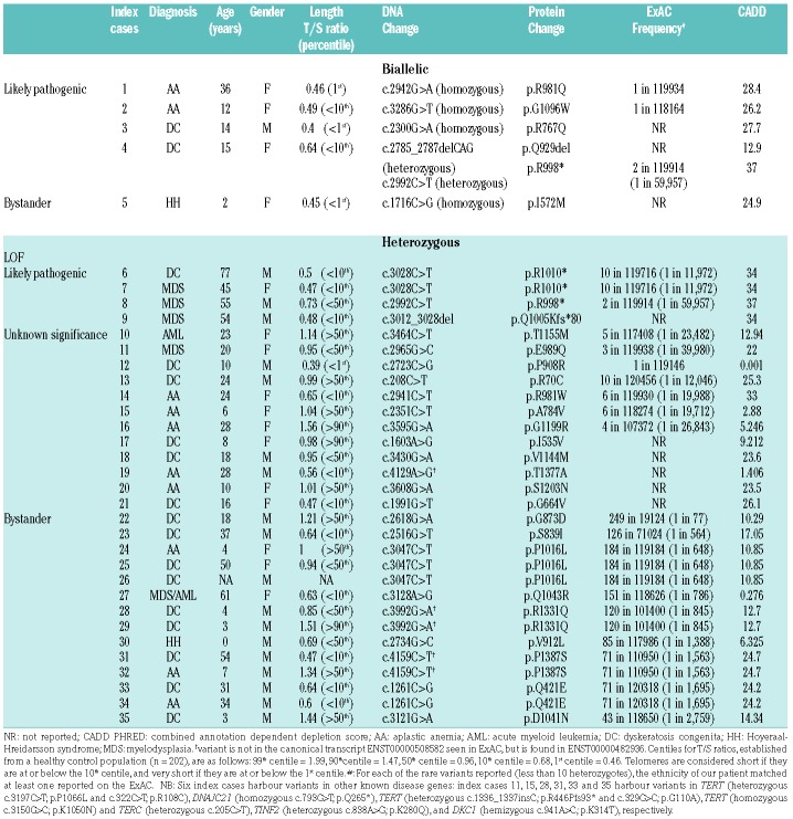
Regulator of telomere elongation helicase 1 (RTEL1) is a DNA helicase involved in telomere maintenance.1,2 Germline biallelic RTEL1 variants have been previously reported in a subset of patients with dyskeratosis congenita (DC) and its severe variant Hoyeraal-Hreidarsson syndrome (HH).3–6 Furthermore, germline heterozygous RTEL1 variants have been linked to a subset of patients with pulmonary fibrosis.2,7,8 We have undertaken sequencing analysis (whole exome and targeted9) of RTEL1, using genomic DNA extracted from peripheral blood of 429 patients from our international bone marrow failure registry which includes DC, HH, aplastic anemia (AA), and familial myelodysplasia/leukemia (MDS/AML). This has revealed that 35 out of the 429 patients have RTEL1 variants (Table 1). Based on the minor allele frequency in the population reported on the Exome Aggregation Consortium database (ExAC – http://exac.broadinstitute.org/), the type of variant (missense, nonsense and indels), telomere length, the Combined Annotation Depletion (CADD) score,10 and segregation as well as information found in literature, we classified these variants into four different groups: (1) biallelic variants, (2) heterozygous loss of function (LOF) variants, (3) heterozygous missense variants of unknown significance (VUS) and (4) heterozygous missense bystander variants.
Table 1.
RTEL1 variants identified in 35 index cases.
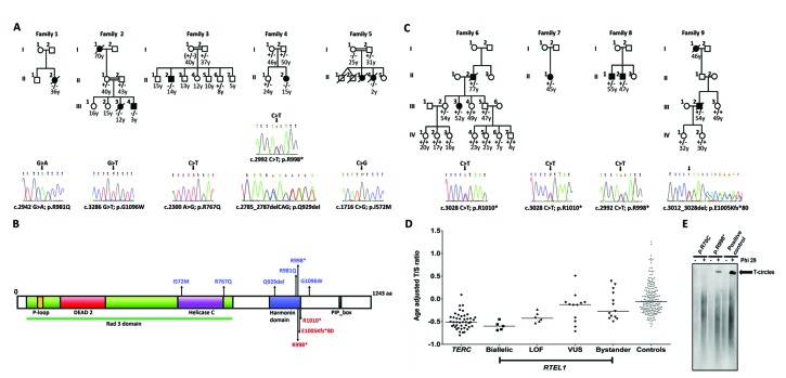
As a result, we have further defined the relationship between variants in the RTEL1 gene and this spectrum of disease. The initial disease association was made when biallelic RTEL1 variants were shown to cause early onset of a severe form of DC and HH.3,5,6,11 Here, we describe five new biallelic families (Figure 1A and 1B), where the variants are believed to be disease-causing in four (Families 1–4). Two families presented with AA, two with DC, and one with HH (Table 1 and Table S1). Interestingly, in one of these the index case presented with AA in adulthood. In the HH family (Family 5), we believe the homozygous RTEL1 variant is a bystander as it did not segregate with disease, being homozygous in both the index case, with severe disease in infancy, and in the asymptomatic 25-year-old mother.
Figure 1.
The segregation, location and impact of RTEL1 variants. (A) Families with biallelic RTEL1 variants and sequencing traces of index cases (homozygous, Families 1, 2, 3 and 5; compound heterozygous, Family 4). The genotyping is described as follows: wild-type (+/+), heterozygous (+/−) or biallelic (−/−). The age at study is given in years. Affected individuals are coloured in black. NA: not available. (B) RTEL1 protein (NP_116575.3) schematic showing the location of the biallelic variants in blue, and the heterozygous LOF variants in red. Conserved protein domains include the P-loop NTPase (yellow); the Rad3 domain (green) which includes the DEAD2 domain (red) and the Helicase C-terminal domain (purple); Harmonin N-like domain (blue); PIP-box – the proliferating cell nuclear antigen interacting protein domain (black). (C) Families with heterozygous LOF RTEL1 variants and sequencing traces of index cases, annotated as in panel A. (D) Age adjusted telomere length values (delta-tel) were measured by subtracting the observed T/S ratio from the expected T/S ratio, using the equation derived from the line of best fit through the plot of T/S ratios from healthy control samples against age. Patients with TERC variants are included as a group with known short telomeres. Centiles were calculated from the control delta-tel values as follows: 99th centile = 0.95, 90th centile = 0.42, 50th centile = 0.06, 10th centile = −0.34, 1st centile = −0.54. The different genotypes are represented as follows, TERC-circles (n=44); biallelic-squares (n=5); loss of function (LOF)-triangles (n=6); variants of unknown significance (VUS)-diamonds (n=12); bystanders-inverted triangles (n=13); controls-grey squares (n=202). (E) T-circle amplification using Phi29 polymerase detected by Southern Blot analysis. Samples: p.R70C - patient with sporadic DC carrying this variant of unknown significance (patient 13 in Table 1); p.R998* - proband of Family 8 carrying this LOF variant (Table 1); positive control - genomic DNA extracted from WI-38 VA-13 cells, known to produce T-circle.
Three disease causing heterozygous LOF RTEL1 variants (nonsense and frameshift deletion) were found in patients from four unrelated families (Figure 1B and 1C), one of whom presented with DC, and the others with MDS and/or liver disease. Therefore, this extends the phenotypes associated with heterozygous loss of function RTEL1 variants to include late onset of MDS and liver disease (Figure 1C and Table 2). This combination of hematological and liver disease is very reminiscent of that established for heterozygous variants in another telomere related gene, TERT,12 which can also present with a severe early onset disease when the variants are biallelic.13
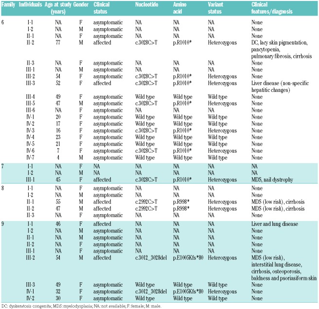
Table 2.
Characteristics of families with RTEL1 LOF variants.
The families we present clearly illustrate the variable penetrance of heterozygous RTEL1 variants. This is exemplified by Family 6 (Figure 1C, p.R1010*) where the index case had DC features, which did not become apparent until age 77 years. His daughter had liver disease at the age of 52 years, and segregation analysis identified four asymptomatic carriers aged below 50 years. This family highlights not only variable penetrance of heterozygous LOF variants, but also suggests a late onset disease predisposition. The same RTEL1 variant was identified in Family 7 (Figure 1C and Table 2), where it was associated with MDS and nail dystrophy in the 45-year-old index case. Interestingly, this same variant is reported by Ballew et al.6 in a heterozygous state as being the cause of HH in two siblings (aged three and one years) with very short telomeres. In the family in question, the mother also harboured the variant, had short telomeres but was asymptomatic. Indeed, in most of the families where the index case has disease due to biallelic RTEL1 variants, both here and in previous reports, the heterozygous parents are generally asymptomatic. However, we now note that these individuals may nevertheless be predisposed to developing disease in their later years. This is suggested by Family 2 (Figure 1A, p.G1096W) where there is a history of pulmonary disease in the grandmother in her 70s and for the p.R998* variant, which has been seen in both severe recessive3,5,6 and late onset dominant settings (Families 4 and 8 and Cogan et al.7). Thus, it is important to be careful when counselling families.
We have identified 14 unrelated patients (Table 1 and Online Supplementary Table S2) with nine heterozygous missense variants we believe to be bystanders due to their occurrence at an allele frequency of more than 1 in 3,000 in the ExAC population. Additionally, 12 unrelated patients with DC (n=5), AA (n=5), MDS (n=1) and AML (n=1) were found to harbour rare heterozygous missense variants. We have classified these as variants of unknown significance (VUS) as they are either not seen in the ExAC population or are present at an allele frequency of less than 1 in 10,000 (Table 1 and Online Supplementary Table S2). The average CADD score for these VUS (average 15.43, range 0.001 – 33), is lower compared to those that we believe to be disease-causing (average 30.13, range 12.9 – 37, Table 1). We also note that of the 15 heterozygous RTEL1 variants previously reported to be associated with pulmonary disease,2,7,8 eight of them are missense.
We have measured telomere lengths by monochrome multiplex quantitative PCR14 in peripheral blood DNA from all patients except for one who had poor DNA quality (Table 1 and Figure 1D). In agreement with previous studies reporting the impact of RTEL1 variants on telomere length, we observed that patients with biallelic variants and those with heterozygous loss of function variants had significantly shorter telomeres than controls, as determined by the age-adjusted T/S ratio (P=0.0005 and P=0.003 respectively, 1-way ANOVA with Dunn’s multiple comparison test). The median age adjusted T/S ratio for the biallelic group is below the 1st centile (−0.6 compared with −0.54), and for the LOF group it is below the 10th centile (−0.43 compared with −0.34). It is interesting to note that for the VUS variants, there appears to be two subgroups. The lower four points correspond to p.G664V, p.P908R, p.R981W and p.T1377A. Three of these variants affect key domains within the protein and may impact on the function of RTEL1 (Figure 1B). These are the helicase C domain (G644V) and the harmonin domain (P908R and R981W).
We previously reported a recurrent missense variant p.R981W as a compound heterozygote in three young unrelated probands (under 12-years-old) with HH.3 Here, we observed the same variant in a heterozygous state in a 24-year-old patient with AA from a consanguineous family (patient 14 in Table 1 and Online Supplementary Table S2). In this case, there is no strong evidence that this variant p.R981W is the cause of AA on account of the relatively high frequency of this variant in the ExAC population (6 in 119930 alleles). However, we do note the short telomeres in this patient and the very high CADD score of this variant, indicating the possibility that it acts as a risk factor for disease.
When a patient presents with an RTEL1 variant, it is difficult to know whether or not it should be considered pathogenic as there are a multitude of rare coding RTEL1 variants in the population at large. Using the ExAC database, the sum of the number of very rare heterozygous coding alleles (at a frequency of <0.0001) is 1,195 in an average of approximately 56,700 people. This is significantly lower than the number of very rare coding variants that we have identified in our cohort (22 in 429 patients, Fisher’s exact test, P = 0.003), but on a case-by-case basis, this background poses a problem. In addition to looking at the ExAC database for population frequency, there are several parameters that we have used to assign pathogenic status. The association of the rare variant with the pathology is a given if the patient under review is presenting with one of the RTEL1 related disease features. Telomere length measurement is now widely used and our experience here is that the heterozygotes, who are often more elderly, may have telomere lengths that are short, but not necessarily very short. We have also looked at T-circles15 and shown that, in some cases, their presence is clearly increased where there is a loss of function variant compared to a common missense variant (Figure 1E). However, this test is not easy to perform, and a normal range has not been established. The in silico prediction tools are helpful and improving, but remain a guide rather than a definitive test. Finally, the segregation of the variant with disease can be decisive. This is more often the case in exclusion rather than inclusion, as we show in Family 5 (Figure 1A and Online Supplementary Table S1).
In summary, our study reports on several important observations. Firstly, heterozygous LOF RTEL1 variants are associated with myelodysplasia and liver disease in adulthood. Secondly, biallelic RTEL1 variants can present with just bone marrow failure in adulthood. Thirdly, many heterozygous variants, and even some biallelic RTEL1 variants, are bystanders. Therefore, in order to assign an accurate status to each RTEL1 variant, detailed clinical and laboratory studies are necessary.
Supplementary Material
Acknowledgments
The authors would like to thank all the clinicians and patients who have helped us over the years, particularly Dr Kiliz. Financial support was provided by The Brazilian National Council for Scientific and Technological Development, Bloodwise, Children with Cancer and the Medical Research Council, UK.
Footnotes
Information on authorship, contributions, and financial & other disclosures was provided by the authors and is available with the online version of this article at www.haematologica.org.
Reference
- 1.Ding H, Schertzer M, Wu X, et al. Regulation of murine telomere length by Rtel: an essential gene encoding a helicase-like protein. Cell. 2004;117(7):873–886. [DOI] [PubMed] [Google Scholar]
- 2.Kannengiesser C, Borie R, Ménard C, et al. Heterozygous RTEL1 mutations are associated with familial pulmonary fibrosis. Eur Respir J. 2015;46(2):474–485. [DOI] [PubMed] [Google Scholar]
- 3.Walne AJ, Vulliamy T, Kirwan M, Plagnol V, Dokal I. Constitutional mutations in RTEL1 cause severe dyskeratosis congenita. Am J Hum Genet. 2013;92(3):448–453. [DOI] [PMC free article] [PubMed] [Google Scholar]
- 4.Jullien L, Kannengiesser C, Kermasson L, et al. Mutations of the RTEL1 helicase in a Hoyeraal-Hreidarsson syndrome patient highlight the importance of the ARCH domain. Hum Mutat. 2016; 37(5):469–472. [DOI] [PubMed] [Google Scholar]
- 5.Deng Z, Glousker G, Molczan A, et al. Inherited mutations in the helicase RTEL1 cause telomere dysfunction and Hoyeraal-Hreidarsson syndrome. Proc Natl Acad Sci USA. 2013; 110(36):E3408–3416. [DOI] [PMC free article] [PubMed] [Google Scholar]
- 6.Ballew BJ, Yeager M, Jacobs K, et al. Germline mutations of regulator of telomere elongation helicase 1, RTEL1, in Dyskeratosis congenita. Hum Genet. 2013;132(4):473–480. [DOI] [PMC free article] [PubMed] [Google Scholar]
- 7.Cogan JD, Kropski JA, Zhao M, et al. Rare variants in RTEL1 are associated with familial interstitial pneumonia. Am J Respir Crit Care Med. 2015;191(6):646–655. [DOI] [PMC free article] [PubMed] [Google Scholar]
- 8.Stuart BD, Choi J, Zaidi S, et al. Exome sequencing links mutations in PARN and RTEL1 with familial pulmonary fibrosis and telomere shortening. Nat Genet. 2015;47(5):512–517. [DOI] [PMC free article] [PubMed] [Google Scholar]
- 9.Walne AJ, Collopy L, Cardoso S, et al. Marked overlap of four genetic syndromes with dyskeratosis congenita confounds clinical diagnosis. Haematologica. 2016;101(10):1180–1189. [DOI] [PMC free article] [PubMed] [Google Scholar]
- 10.Kircher M, Witten DM, Jain P, O’Roak BJ, Cooper GM, Shendure J. A general framework for estimating the relative pathogenicity of human genetic variants. Nat Genet. 2014;46(3):310–315. [DOI] [PMC free article] [PubMed] [Google Scholar]
- 11.Le Guen T, Jullien L, Touzot F, et al. Human RTEL1 deficiency causes Hoyeraal-Hreidarsson syndrome with short telomeres and genome instability. Hum Mol Genet. 2013;22(16):3239–3249. [DOI] [PubMed] [Google Scholar]
- 12.Calado RT, Regal JA, Kleiner DE, et al. A spectrum of severe familial liver disorders associate with telomerase mutations. PLoS One. 2009;4(11):e7926. [DOI] [PMC free article] [PubMed] [Google Scholar]
- 13.Marrone A, Walne A, Tamary H, et al. Telomerase reverse-transcriptase homozygous mutations in autosomal recessive dyskeratosis congenita and Hoyeraal-Hreidarsson syndrome. Blood. 2007;110(13):4198–5205. [DOI] [PMC free article] [PubMed] [Google Scholar]
- 14.Cawthon RM. Telomere length measurement by a novel monochrome multiplex quantitative PCR method. Nucleic Acids Res. 2009;37(3):e21. [DOI] [PMC free article] [PubMed] [Google Scholar]
- 15.Zellinger B, Akimcheva S, Puizina J, et al. Ku suppresses formation of telomeric circles and alternative telomere lengthening in Arabidopsis. Mol Cell. 2007;27(1):163–169 [DOI] [PubMed] [Google Scholar]
Associated Data
This section collects any data citations, data availability statements, or supplementary materials included in this article.


