Abstract
The metabolic enzymes that compose glycolysis, the citric acid cycle, and other pathways within central carbon metabolism have emerged as key regulators of animal development. These enzymes not only generate the energy and biosynthetic precursors required to support cell proliferation and differentiation, but also moonlight as regulators of transcription, translation, and signal transduction. Many of the genes associated with animal metabolism, however, have never been analyzed in a developmental context, thus highlighting how little is known about the intersection of metabolism and development. Here we address this deficiency by using the Drosophila TRiP RNAi collection to disrupt the expression of over 1,100 metabolism-associated genes within cells of the eye imaginal disc. Our screen not only confirmed previous observations that oxidative phosphorylation serves a critical role in the developing eye, but also implicated a host of other metabolic enzymes in the growth and differentiation of this organ. Notably, our analysis revealed a requirement for glutamine and glutamate metabolic processes in eye development, thereby revealing a role of these amino acids in promoting Drosophila tissue growth. Overall, our analysis highlights how the Drosophila eye can serve as a powerful tool for dissecting the relationship between development and metabolism.
Keywords: Drosophila, metabolism, mitochondria, oxidative phosphorylation, glutamine metabolism
The fruit fly Drosophila melanogaster has emerged as a powerful model for investigating the metabolic mechanisms that support animal growth and development. In this regard, a key advantage of studying metabolism in the fly is that the disruption of an individual metabolic reaction often induces a specific phenotype, thus revealing energetic and biosynthetic bottlenecks that influence cell growth, proliferation, and differentiation. For example, mutations that disrupt activity of the citric acid cycle (TCA cycle) enzymes Isocitrate Dehydrogenase 3b (Idh3b) and Malate Dehydrogenase 2 (Mdh2) prevent the larval salivary glands from dying at the onset of metamorphosis (Wang et al. 2008; Wang et al. 2010; Duncan et al. 2017). These observations suggest that the salivary glands are uniquely dependent on the TCA cycle to activate the cell death program and reveal an unexpected relationship between central carbon metabolism and metamorphosis. Such phenotype-driven studies are essential for investigating how metabolism and development are coordinated during the fly life cycle.
The Drosophila eye has long served as a powerful model for both metabolism and development (for reviews, see Dickinson and Sullivan 1975; Kumar 2018). Many of the earliest genetic studies conducted in the fly were based upon genes such as vermillion, cinnabar, and rosy, which control eye pigmentation and encode enzymes involved in tryptophan and purine metabolism (Lindsley and Zimm 1992). Similarly, classic work by Beadle and Ephrusi used transplantation experiments to demonstrate that ommochromes are synthesized in larval peripheral tissues and transported into the eye (Beadle and Ephrussi 1936), thus revealing that metabolism is systemically coordinated during development. Nearly a century later, the Drosophila eye still serves as an essential tool for studying developmental metabolism – a fact that is best illustrated by a finding from Utpal Banerjee’s lab. In a classic demonstration of how unbiased screens can identify unexpected developmental regulators, members of the Banerjee lab discovered that the Drosophila gene CoVa (FBgn0019624; also known as tenured and COX5A), a subunit of Complex IV within the electron transport chain (ETC), is essential for normal eye development (Mandal et al. 2005). While such a discovery could have been easily discounted as the disruption of a housekeeping gene, characterization of CoVa mutants demonstrated that reduced oxidative phosphorylation (OXPHOS) induces a G1 cell-cycle arrest during the second mitotic wave (Mandal et al. 2005). Moreover, this phenomenon was found to be orchestrated by the metabolic sensor AMPK, which responds to the decreased ATP levels present within CoVa mutant cells by activating p53 and lowering Cyclin E levels (Mandal et al. 2005). These studies of CoVa function, together with similar studies of other electron transport chain (ETC) subunits and mitochondrial tRNAs (Mandal et al. 2005; Liao et al. 2006), demonstrate that the eye can be used to efficiently understand how metabolism is integrated with developmental signaling pathways.
Here we use the Drosophila TRiP RNAi collection to identify additional metabolism-associated genes that influence eye development. Our screen used the eyes absent composite enhancer-GAL4 (eya composite-GAL4) driver to induce expression of 1575 TRiP RNAi transgenes (representing 1129 genes) during development of the eye imaginal disc (Weasner et al. 2016). This analysis not only confirmed previous findings that genes involved in OXPHOS are essential for eye development, but also uncovered a role for glutamate and glutamine metabolism within this tissue. Moreover, we identified several poorly characterized enzymes that are essential for normal eye formation, thus hinting at novel links between metabolism and tissue development. Overall, our genetic screen provides a snapshot of the biosynthetic and energetic demands that the development of a specific organ imposes upon intermediary metabolism.
Materials and Methods
Drosophila Strains and Husbandry
Fly stocks and crosses were maintained at 25° on Bloomington Drosophila Stock Center (BDSC) food. All genetic crosses described herein used eya composite-GAL4 to induce transgene expression (a kind gift from Justin Kumar’s lab, Weasner et al. 2016). The TRiP RNAi lines used in this study were selected by searching the BDSC stock collection using a previously described list of metabolism-associated genes (Tennessen et al. 2014; Perkins et al. 2015). All strains used in this study are available through the BDSC.
Genetics Crosses and Phenotypic Characterization
Five adult male flies from each TRiP stock was crossed with five w1118; eya composite-GAL4 adult virgin females. For each cross, F1 progeny heterozygous for both eya composite-GAL4 and the UAS-RNAi transgene were scored for eye phenotypes within three days of eclosion. Eyes were scored for the following phenotypes: rough, glossy, small, no eye, misshaped, overgrowth, necrosis, abnormal pigmentation, and lethality prior to eclosion. Whenever possible, at least 20 adults were scored from during this screen. Any TRiP stock that produced a phenotype during the initial analysis was reanalyzed using the same mating scheme described above and twenty flies of each sex were scored. In some instances, expression of the TRiP transgene induced a lethal or semi-lethal phenotype prior to eclosion, thus limiting the number of animals that could be scored in our analysis. To avoid confirmation bias, each cross was only labeled with the BDSC strain number and the genotype was revealed only after phenotypic characterization.
Statistical Analysis
Genes were assigned to individual pathways based on the metabolic pathways annotated within the Kyoto Encyclopedia of Genes and Genomes (KEGG) pathways (Kanehisa 2017; Kanehisa et al. 2019). Enrichment analyses for individual metabolic pathways were conducted using Fisher’s exact test with the P value being calculated using two tails. For the purpose of these calculations, expression of 165 transgenes induced a phenotype while the expression of 1410 transgenes had no effect on eye development.
Data Availability
All TRiP lines used in this study are available from the BDSC. The eya composite-GAL4 strain is available upon request. Data generated in this study have been uploaded to the RNAi Stock Validation and Phenotype (RSVP) database, which is publicly accessible through the DRSC/TRiP Functional Genomics Resources website. The full list of TRiP stocks used in our analysis can be found in Table S2 and the strains analyzed in the secondary screen are listed in Table S3. To facilitate replication of our study, all supplemental tables contain both the BDSC and FlyBase identification numbers (Fbgn). Figure S1 includes a schematic diagram of glycolysis and provides a summary of how the TRiP RNAi lines that target this pathway disrupt eye development. Figure S2 includes a schematic diagram of the TCA cycle and illustrates of how TRiP RNAi lines that target this pathway do not disrupt eye development. Table S1 provides the list of Drosophila genes involved in metabolism and nutrient sensing that were used to select the TRiP strains used in this study. Table S2 contains a list of the TRiP RNAi stocks used in this study and the eye phenotype associated with each strain. Table S3 contains the list of TRiP RNAi stocks that induced an eye phenotype during the initial screen and the phenotypes that were observed when these strains were reanalyzed. Table S4 contains the list of TRiP RNAi strains that were used to disrupt oxidative phosphorylation and the eye phenotype induced by each transgene. Table S5 contains the list of TRiP RNAi strains that were used to disrupt GPI anchor biosynthesis and the eye phenotype induced by each transgene. Table S6 contains the list of TRiP RNAi strains that were used to disrupt glycolysis and the eye phenotype induced by each transgene. Table S7 contains the list of TRiP RNAi strains that were used to disrupt the TCA cycle and the eye phenotype induced by each transgene. Table S8 contains the list of TRiP RNAi strains that were used to disrupt glutamate and glutamine metabolism and the eye phenotype induced by each transgene. Supplemental material available at FigShare: https://doi.org/10.25387/g3.8038940.
Results
To identify metabolic processes involved in eye development, we used the eya composite-GAL4 driver to express TRiP RNAi transgenes that target metabolism-associated genes (Table S1). Since this GAL4 driver promotes transgene expression in the eye imaginal disc from the L2 stage until after the morphogenetic furrow moves across the eye field (Weasner et al. 2016), our screen of 1575 TRiP RNAi lines was designed to identify metabolic processes required for the proliferation and differentiation of cells within this organ. Of the RNAi transgenes examined, 198 induced an eye phenotype in the initial screen and 165 subsequently generated reproducible phenotypes (Tables S2 and S3).
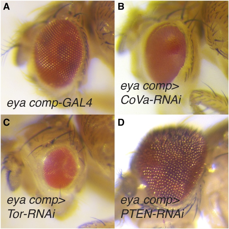
Among the TRiP lines that consistently induced an eye phenotype were a number of positive controls. Notably, the RNAi transgene that targets CoVa (BDSC 27548) induced glossy-eye phenotype (Figure 1A-B; Tables S2 and S3), thus phenocopying the eye defect associated with CoVa mutant clones (Mandal et al. 2005). Our screen also included TRiP lines that targeted components of the insulin and Tor signaling pathways, which modulate developmental growth in response to sugar and amino acid availability (Tennessen and Thummel 2011). As expected, RNAi transgenes targeting positive regulators of these pathways, including the Insulin Receptor (InR; BDSC 35251; FBgn0283499, REF), Phosphatidylinositol 3-kinase (Pi3K92E; BDSC 27690; FBgn0015279), Akt1 (BDSC 31701 and 33615; FBgn0010379), Target of rapamycin (Tor; BDSC 34639; FBgn0021796), and raptor (BDSC 31528, 31529, and 34814; FBgn0029840), resulted in either small or misshapen eyes (Figure 1C; Tables S2 and S3). Similarly, RNAi-induced depletion of the negative growth regulators Phosphatase and tensin homolog (PTEN; BDSC 25967 and 33643; FBgn0026379), Tsc1 (BDSC 52931 and 54034; FBgn0026317) and Tsc2 (BDSC 34737; FBgn0005198) induced an overgrowth phenotype (Figure 1D; Tables S2 and S3). Our findings are consistent with previous studies that described roles for these insulin and Tor signaling pathway components in eye development (Chen et al. 1996; Böohni et al. 1999; Goberdhan et al. 1999; Huang et al. 1999; Ito and Rubin 1999; Verdu et al. 1999; Weinkove et al. 1999; Gao et al. 2000; Oldham et al. 2000; Potter et al. 2001).
Figure 1.
Eye phenotypes caused by RNAi disruption of OXPHOS and growth control. (A) An eya composite-GAL4/+ control eye (eya comp). (B) RNAi depletion of CoVa, targeted using BDSC 27548, resulted in a glossy-eyed phenotype. (C-D) TRiP RNAi transgenes targeting the growth control regulators (C) Tor, targeted using BDSC 33951, and (D) PTEN, targeted using BDSC 25967, induced small and large eyes, respectively. For (B-D), eya composite-GAL4 is abbreviated eya comp.
Our ability to identify TRiP lines that interfere with the expression of known growth regulators suggests that our screen efficiently identified key metabolism-associated genes involved in eye development. We would note, however, that a screen of this nature will inevitably produce false-positive results due to off-target RNAi effects and false negative results due to inefficient depletion of target transcripts. Therefore, we will limit the Results and Discussion sections to those pathways that are either represented by multiple positive results or are notably absent in our analysis.
Oxidative Phosphorylation
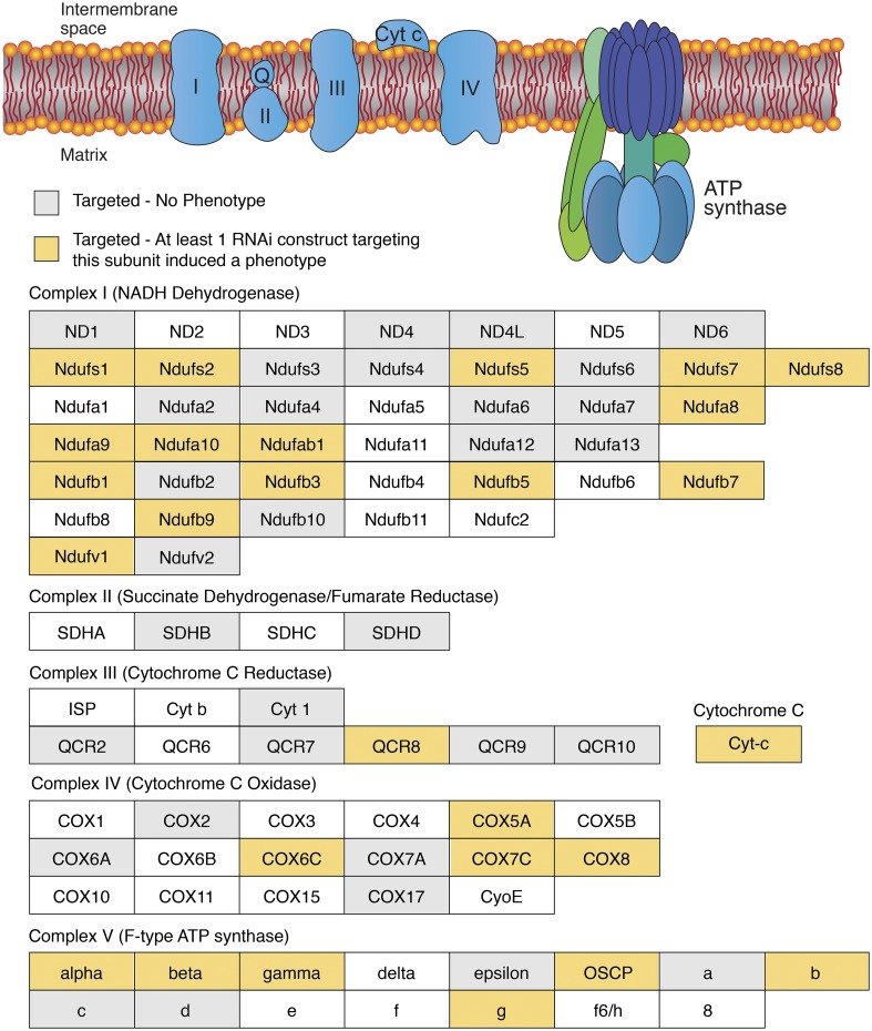
Of the 165 RNAi transgenes that consistently induced an eye phenotype when crossed to the eya composite-GAL4 driver, 40 targeted genes that encode subunits of the ETC and ATP synthase (F-type) as defined by KEGG pathway dme00190 (Figure 2; Table S4). These results indicate that eye development is quite sensitive to disruption of Complex I, Complex IV, and Complex V (F-type ATP-synthase), as nearly half of the transgenes that targeted these complexes induced an eye phenotype (Figure 2; Tables S2 and S3). In addition, expression of the siRNAs that targeted Cytochrome C proximal (Cyt-c-p; FBgn0284248; BDSC 64898) and Coenzyme Q biosynthesis protein 2 (Coq2; FBgn0037574; BDSC 53276), which is required for Coenzyme Q production, resulted in highly penetrant glossy-eye phenotypes (Figure 2; Tables S2-S4). We would also note that while expression of only one siRNA associated with Complex II or III induced a phenotype, our screen included relatively few strains that targeted these complexes.
Figure 2.
The ETC and ATP synthase are required for normal eye development. (Top) A diagram illustrating the ETC and ATP synthase within the inner mitochondrial membrane. (Below) Individual subunits are listed in boxes and organized by complex. Yellow-shaded boxes indicate that at least one RNAi transgene targeting the subunit induced a phenotype. Gray-shaded boxes indicate that none of the RNAi transgenes targeting this subunit induced a phenotype. Corresponding data can be found in Table S4. Diagram is a modified from the illustration presented on the KEGG website for pathway dme00190.
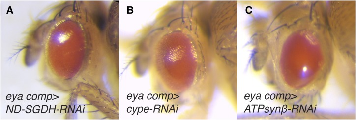
Our findings regarding the ETC and ATP synthase are notable because, among the metabolism-associated TRiP transgenes capable of inducing an eye phenotype, those that disrupt OXPHOS represent one of the largest and most significantly enriched groups (P < 0.0001, Fisher’s exact test, two-tailed, 94 OXPHOS transgenes tested). Moreover, ETC-related transgenes were uniquely associated with the same morphological phenotype - not only did disruption of OXPHOS almost invariably induce a glossy-eye (Figure 3A-C; Table S4), but among the RNAi lines tested, almost all of the transgenes that resulted in a glossy-eye phenotype were associated with OXPHOS (Tables S2 and S3). We would note, however, that there is variability among the TRiP lines regarding the penetrance and severity of the glossy-eye phenotype (compare Figure 3A, 3B, and 3C). This variability could be due to several factors, including RNAi efficacy, the differential requirements for each OXPHOS subunit in promoting ATP production, and unique requirements for individual OXPHOS subunits during eye development. Regardless, the phenotypic similarities displayed among the OXPHOS-associated TRiP lines support two previously stated hypotheses (see Mandal et al. 2005; Liao et al. 2006): (1) ETC subunits influence eye development in a similar manner. (2) Considering that the function of many nuclear-encoded mitochondrial proteins remain unknown (Pagliarini et al. 2008; Pagliarini and Rutter 2013; Calvo et al. 2016), targeted disruption of these uncharacterized genes within the eye imaginal disc could potentially identify novel OXPHOS regulators by simply using the glossy-eye phenotype as a readout.
Figure 3.
Disruption of the ETC and ATP synthase induces a glossy-eye phenotype. Representative images illustrating how RNAi depletion of OXPHOS components induce a glossy-eyed phenotype. (A) ND-SGDH, targeted using BDSC 67311. (B) cype, targeted using BDSC 33878. (C) ATPsynβ, targeted using BDSC 27712. For all images, eya composite-GAL4 is abbreviated eya comp. Please note that the phenotype in these panels is similar to the CoVa-RNAi eye phenotype in Figure 1B. Any perceived differences between (A-C) and Figure 1B is the result of different microscope light sources.
Glycosylphosphatidylinositol (GPI)-Anchor Synthesis
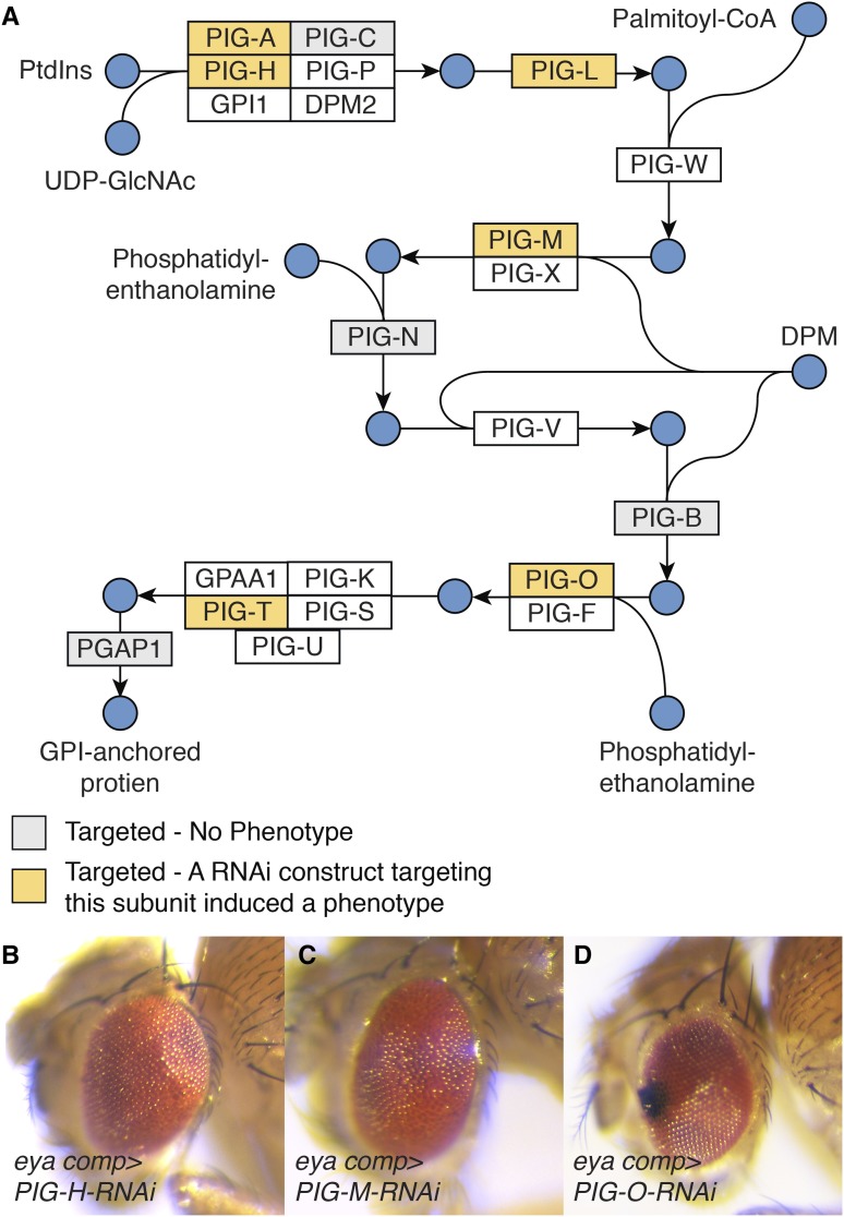
Many of the enzymes involved in GPI-anchor synthesis emerged as being essential for normal eye development. Of the 8 genes that are associated with this metabolic pathway (KEGG dme00563) and were examined during our screen, TRiP lines that targeted five of these genes induced rough eye phenotypes (Figure 4A-D; Table S5). These enzymes represent multiple steps within GPI-anchor biosynthesis, which is consistent with previous observations that this pathway is essential for the function of key proteins involved in eye development, including rhodopsin, chaoptin, and dally (Krantz and Zipursky 1990; Kumar and Ready 1995; Nakato et al. 1995; Satoh et al. 2013). Considering that the GPI-anchor biosynthetic enzymes would be predicted to emerge from a screen of this nature, these findings suggest that our approach effectively identified genes involved in eye development.
Figure 4.
Disruption of GPI-anchor biosynthesis induces a rough eye phenotype. (A) A diagram illustrating GPI-anchor biosynthesis. Diagram is based upon KEGG pathway dme00563. Abbreviations: Phosphatidyl-1D-myo-inositol (PtdIns); Dolichyl phosphate D-mannose (DPM). (B-D) Representative images showing the rough eye phenotype caused by RNAi-induced disruption of (B) PIG-H, targeted using BDSC 67330, (C) PIG-M, targeted using BDSC 38321, and (D) PIG-O, targeted using BDSC 67247. For all images, eya composite-GAL4 is abbreviated eya comp.
Glycolysis and the TCA cycle
Expression of RNAi constructs targeting either glycolysis (KEGG dme00010) or the TCA cycle (KEGG dme00020) rarely affected eye development. Only two of the 40 TRiP lines that disrupt expression of glycolytic enzymes and none of the transgenes that targeted genes associated with the TCA cycle (n = 27) induced an eye phenotype (Figure S1A, S2, and Tables S6 and S7). These results, while surprising, require confirmation using null alleles of these genes, as we can’t eliminate the possibility that enzymes in glycolysis and the TCA cycle are so abundant that RNAi is incapable of reducing their expression below a threshold level. However, we would note that the eyes of Mitochondrial Pyruvate Carrier 1 (Mpc1; FBgn0038662) mutants appear morphologically normal (Figure S1B, Bricker et al. 2012). Considering that Mpc1 mutants are unable to transport pyruvate into their mitochondria, eye development must be able proceed normally when glycolysis is uncoupled from the TCA cycle (Bricker et al. 2012). Second, we previously demonstrated that the TRiP line targeting Phosphofructokinase (Pfk; FBgn0003071; BDSC 34366) reduces Pfk mRNA levels by ∼80%, significantly decreases pyruvate levels, and restricts larval growth (Li et al. 2018), however, Pfk-RNAi does not interfere with eye development (Figure S1C). Although we have not yet confirmed the effectiveness of this Pfk-RNAi transgene in the eye imaginal disc, the absence of a phenotype in our screen is notable and warrants future analysis using Pfk loss-of-function mutations.
While additional studies are required to understand how glycolysis and the TCA cycle influence eye development, and we doubt that either pathway is completely dispensable in this context, our results raise several intriguing hypotheses. Metabolomic studies of the Mpc1 mutants reveal that fly larvae raised on standard media readily adapt to this severe disruption of central carbon metabolism (Bricker et al. 2012). The same compensatory mechanisms that are activated in Mpc1 mutants could also support eye development under conditions of reduced glycolytic flux. In addition, considering the apparent dependence of developing eye cells on catabolism of the amino acid glutamine (see below), glucose might not be the primary energy source used by these cells. Finally, we would note that when compared with other larval organs, such as the muscle and brain, imaginal discs express low levels of Lactate Dehydrogenase (dLdh, Rechsteiner 1970; Wang et al. 2016). Therefore, glycolytic flux appears to be regulated differently in the eye when compared with other larval tissues.
Pentose Phosphate Pathway
Two enzymes within the oxidative shunt of the pentose phosphate pathway (KEGG dme0030), glucose-6-phosphate dehydrogenase (G6PDH; known as Zwischenferment; FBgn0004057; BDSC 50667) and phosphogluconate dehydrogenase (Pgd; FBgn0004654; BDSC 65078) produced a small eye phenotype (Tables S2 and S3). These results were unexpected because both enzymes are thought to be dispensable for growth and viability - Zw mutants display no discernable phenotype, and while Pgd mutants are lethal, Zw Pgd double mutant are viable with no obvious morphological defects (Hughes and Lucchesi 1977). While such results require confirmation using clonal analysis, our observations hint at the possibility that tissue-specific disruption of the pentose phosphate pathway can induce developmental phenotypes – a phenomenon that has been previously observed in studies of Drosophila metabolism (Caceres et al. 2011). Considering that the oxidative branch of the pentose phosphate pathway serves a key role in maintaining NAPDH levels (Ying 2008), future studies should examine the possibility that eye development relies on G6PDH and PGD to maintain this pool of reducing equivalents.
Glutamine metabolism
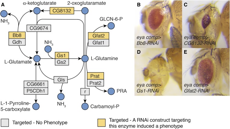
Our screen revealed an unexpected role for glutamine (Gln) and glutamate (Glu) in eye development. Of the 24 TRiP lines that targeted genes directly involved in Gln/Glu metabolism (see enzymes that interact with Gln/Glu in KEGG pathway dme00250), five induced either a small or no eye phenotype (Table S8). These five RNAi lines targeted five genes that directly regulate Gln/Glu-dependent metabolic processes (Figure 5A):
big bubble 8 (bb8; FBgn0039071; BDSC 57484) encodes the enzyme glutamate dehydrogenase (GLUD), which is responsible for converting glutamate into α-ketoglutarate and ammonia (see KEGG dme00250). Because GLUD can funnel glutamate into the TCA cycle, this enzyme allows cells to generate both fatty acids and ATP in a glucose-independent manner – as is evident by the fact that many cancer cells adapt to inhibition of glycolysis by up-regulating GLUD activity (Altman et al. 2016). A recent study in Drosophila has implicated bb8 in promoting spermatogenesis (Vedelek et al. 2016).
CG8132 (FBgn0037687; BDSC 57794) encodes an omega-amidase that is homologous to the human nitrilase family member 2 (NIT2) enzyme, which converts α-ketoglutaramate into a-ketoglutarate and ammonia (Jaisson et al. 2009; Krasnikov et al. 2009). The endogenous function of this enzyme remains poorly understood in animal systems, however, there are some indications that NIT2 functions as a tumor suppressor in humans (Zheng et al. 2015).
Glutamine synthetase 1 (Gs1; FBgn0001142; BDSC 40836) encodes an enzyme that generates Gln from ammonia and Glu (Caizzi and Ritossa 1983). Since Gln is required for several biosynthetic processes, including the production of nucleotides, glutathione, and glucosamine-6-phosphate (see below, Altman et al. 2016), Gs1 ensures that growing and proliferating cells have adequate levels of this amino acid. In Drosophila, this enzyme is also required for early mitotic cycles within syncytial embryos (Frenz and Glover 1996).
Glutamine:fructose-6-phosphate aminotransferase 2 (Gfat2; FBgn0039580; BDSC 34740) encodes an enzyme that converts Gln and fructose-6-phosphate into Glu and glucosamine-6-phosphate (Graack et al. 2001). In turn, glucosamine-6-phosphate is used to generate N-acetyl-glucosamine (GlcNAc), which is required for several cellular processes, including chitin formation and protein modifications. Moreover, the multifaceted roles for glucosamine-6-phosphate and GlcNAc in development are essential for cell proliferation and tissue growth, as demonstrated by the recent observation that Drosophila Gfat2 is required for proliferation of adult intestinal stem cells (Mattila et al. 2018).
phosphoribosylamidotransferase (Prat; FBgn0004901; BDSC 43296) links Gln with purine biosynthesis (Clark 1994). RNAi targeting of this gene in the eye imaginal disc resulted in a lethal phenotype during our initial screen (Table S2), suggesting that disruption of nucleotide production in the developing eye induces non-autonomous effects. Our observation is consistent with previous results, which indicate that Prat is expressed in L3 imaginal discs and that Prat-RNAi results in a pupal lethality (Ji and Clark 2006; Brown et al. 2014).
In addition to the enzymes that are directly involved in Glu/Gln catabolism, RNAi of two additional genes associated with these amino acids elicited eye phenotypes:
Figure 5.
Enzymes associated with glutamate (Glu) and glutamine (Gln) metabolism are essential for normal eye development. (A) A diagram illustrating the metabolic reactions associated with Glu and Gln metabolism as defined by KEGG pathway dme00250. (B-E) Representative images illustrating how disruption of Glu/Gln metabolism affects eye development. Abbreviations: D-glucosamine-6-phosphate (GLCN-6-P) and 5-phosphoribosylamine (PRA). (B) Bb8, targeted using BDSC 57484. (C) CG8132, targeted using BDSC 38321. (D) Gs1, targeted using BDSC 40836. (E) Gfat2, targeted using BDSC 34740. For all images, eya composite-GAL4 is abbreviated eya comp.
γ-glutamyl transpeptidase (Ggt-1; FBgn0030932; BDSC 64529) encodes an enzyme that transfers a γ-glutamyl residue from a donor molecule, such as glutathione, to an acceptor molecule (Ikeda and Taniguchi 2005; Heisterkamp et al. 2008). Moreover, this enzyme can generate Glu by using water as an acceptor molecule for γ-glutamyl (Ikeda and Taniguchi 2005; Heisterkamp et al. 2008). Drosophila Ggt-1 was previously reported to function in the larval Malpighian tubules, where it facilitates green-light avoidance by generating glutamate (Liu et al. 2014).
Selenide water dikinase (SelD; FBgn0261270; BDSC 29553) encodes a member of the selenophosphate synthetase 1 (SPS1) enzyme family that does not synthesize selenophosphate but rather functions in redox homeostasis and glutathione metabolism (Xu et al. 2007a; Xu et al. 2007b; Tobe et al. 2016). Consistent with the proposed functions of SPS1 proteins, SelD serves a critical role in Drosophila eye development by restricting reactive oxygen species (ROS) accumulation (Morey et al. 2003). In the absence of SelD function, elevated ROS levels within the eye interfere with a variety of developmental signaling events (Alsina et al. 1999; Morey et al. 2001), as evident by the fact that the SelD null mutation patufet (SelDptuf) dominantly suppresses the eye and wing phenotypes induced by ectopic activation of sevenless and Raf (Morey et al. 2001). While the exact metabolic function of SelD remains unknown, SelD knockdown in SL2 cells induces excessive Gln accumulation (Shim et al. 2009).
We find these results notable because these seven enzymes are involved in a diversity of metabolic processes, including biosynthesis, energy production, and cell signaling. Not only are many of these enzymes implicated in cancer cell proliferation and tumor growth (Lin et al. 2007; Altman et al. 2016), but one of the metabolites associated with these enzymes, α-ketoglutarate, is an essential regulator of histone methylation and gene expression (Chisolm and Weinmann 2018). Moreover, since both glutamine and α-ketoglutarate were recently found to activate Drosophila Tor (Zhai et al. 2015; Yoon et al. 2017), disruption of Glu/Gln metabolism could affect eye development by restricting Tor signaling. Overall, our findings indicate that Drosophila eye development could serve as a powerful in vivo model for investigating how Glu/Gln metabolism influences cell proliferation and tissue growth.
Discussion
Here we use the Drosophila TRiP RNAi collection to identify metabolic processes that are required for the growth and development of the eye. Our screen not only verified that RNAi could effectively disrupt metabolic processes with known roles in eye development (e.g., CoVa, ETC subunits, enzymes involved in GPI-anchor biosynthesis), but also proved effective at identifying additional pathways that are essential for the growth of this tissue. Here we highlight two key findings that we believe warrant further examination.
Metabolic pathways are associated with specific developmental events
The RNAi phenotypes uncovered in our screen demonstrate how different stages in eye development impart unique demands on intermediary metabolism. For example (and as previously described by the Banerjee lab), the OXPHOS-associated glossy eye phenotype results from a cell cycle arrest during the second mitotic wave (Mandal et al. 2005), resulting in the loss of pigment cells and lens secreting cone cells (for a review of cone and pigment cell development, see Kumar 2012). A key feature of this phenotype is that the overall eye size remains normal, indicating that OXPHOS disruption does not interfere with cell proliferation ahead of the morphogenetic furrow. The unique nature of this phenotype suggests that any TRiP line inducing a glossy, normal sized eye should be investigated for a potential role in OXPHOS. Similarly, the rough eye phenotype induced by RNAi of GPI-anchor biosynthesis likely reflects the disruption of proteins required for the formation of ommatidium, including those associated with morphogen signaling, cell polarity, and cell specification (Kumar 2012). Therefore, those genes associated with a rough eye phenotype in our screen should be examined for potential roles in ommatidial assembly.
While our screen indicates that dozens of metabolic enzymes are required for eye development, perhaps our most intriguing results are the small/no eye phenotypes induced by the disruption of Glu/Gln metabolism. These developmental defects likely stem from either decreased cell proliferation ahead of the morphogenetic furrow or defects in cell fate specification (for review, see Kumar 2011) and are consistent with the role of Glu/Gln-associated enzymes in mammalian cell proliferation and differentiation (Altman et al. 2016). The developing eye disc, therefore, provides an ideal model to understand how signal transduction cascades regulate Glu/Gln metabolism and investigate how the metabolism of these amino acids influence cell proliferation and tissue growth.
The Drosophila eye as a model for studying metabolic plasticity and robustness
Our screen further supports previous observations that Drosophila development is surprisingly resistant to metabolic insults. The observation that eye development was largely unaffected by the RNAi transgenes that target glycolysis and the TCA cycle was unexpected. While we doubt that either pathway is completely dispensable for eye formation, our results are consistent with the ability of Drosophila development to withstand severe metabolic insults (e.g., Mpc1 mutants, Bricker et al. 2012). This metabolic robustness makes sense because animal development must readily adapt to a variety of nutrient sources and environmental stresses. Based upon the results of this screen, we propose that the fly eye could serve as a model to identify the compensatory pathways that that allow cell growth and proliferation to proceed in the face of major metabolic disruptions.
Overall, our genetic screen demonstrates how Drosophila melanogaster can serve as a powerful model to identify tissue-specific metabolic factors required for tissue growth and organogenesis. Moreover, we believe this work represents a necessary step toward systematically analyzing the metabolic pathways that support cell proliferation and tissue growth within the fly.
Acknowledgments
We thank the TRiP at Harvard Medical School (NIH/NIGMS R01-GM084947) for providing transgenic RNAi fly stocks used in this study. We also thank the Bloomington Drosophila Stock Center (NIH P40OD018537), Cale Whitworth for helping us search the BDSC database, Flybase (NIH 5U41HG000739), and the members of the Kumar lab for providing reagents and technical support. Thanks to Kudakawashe Tshililiwa, Curteisha Jacobs, and Joy Morounfolu for assistance with genetic crosses. Special thanks to Bonnie Weasner and Justin Kumar for support, advice, helpful discussions, and a critical reading of the mansucript. J.M.T. is supported by the National Institute of General Medical Sciences of the National Institutes of Health under a R35 Maximizing Investigators’ Research Award (MIRA; 1R35GM119557).
Footnotes
Supplemental material available at FigShare: https://doi.org/10.25387/g3.8038940.
Communicating editor: A. Bashirullah
Literature Cited
- Alsina B., Corominas M., Berry M. J., Baguna J., Serras F., 1999. Disruption of selenoprotein biosynthesis affects cell proliferation in the imaginal discs and brain of Drosophila melanogaster. J. Cell Sci. 112: 2875–2884. [DOI] [PubMed] [Google Scholar]
- Altman B. J., Stine Z. E., Dang C. V., 2016. From Krebs to clinic: glutamine metabolism to cancer therapy. Nat. Rev. Cancer 16: 619–634 (errata: Nat. Rev. Cancer 16: 749, 773). 10.1038/nrc.2016.71 [DOI] [PubMed] [Google Scholar]
- Beadle G. W., Ephrussi B., 1936. The Differentiation of Eye Pigments in Drosophila as Studied by Transplantation. Genetics 21: 225–247. [DOI] [PMC free article] [PubMed] [Google Scholar]
- Böhni R., Riesgo-Escovar J., Oldham S., Brogiolo W., Stocker H., et al. , 1999. Autonomous control of cell and organ size by CHICO, a Drosophila homolog of vertebrate IRS1–4. Cell 97: 865–875. 10.1016/S0092-8674(00)80799-0 [DOI] [PubMed] [Google Scholar]
- Bricker D. K., Taylor E. B., Schell J. C., Orsak T., Boutron A., et al. , 2012. A mitochondrial pyruvate carrier required for pyruvate uptake in yeast, Drosophila, and humans. Science 337: 96–100. 10.1126/science.1218099 [DOI] [PMC free article] [PubMed] [Google Scholar]
- Brown J. B., Boley N., Eisman R., May G. E., Stoiber M. H., et al. , 2014. Diversity and dynamics of the Drosophila transcriptome. Nature 512: 393–399. 10.1038/nature12962 [DOI] [PMC free article] [PubMed] [Google Scholar]
- Caceres L., Necakov A. S., Schwartz C., Kimber S., Roberts I. J., et al. , 2011. Nitric oxide coordinates metabolism, growth, and development via the nuclear receptor E75. Genes Dev. 25: 1476–1485. 10.1101/gad.2064111 [DOI] [PMC free article] [PubMed] [Google Scholar]
- Caizzi R., Ritossa F., 1983. The enzyme glutamine synthetase I of Drosophila melanogaster is associated with a modified RNA. Biochem. Genet. 21: 267–285. 10.1007/BF00499138 [DOI] [PubMed] [Google Scholar]
- Calvo S. E., Clauser K. R., Mootha V. K., 2016. MitoCarta2.0: an updated inventory of mammalian mitochondrial proteins. Nucleic Acids Res. 44: D1251–D1257. 10.1093/nar/gkv1003 [DOI] [PMC free article] [PubMed] [Google Scholar]
- Chen C., Jack J., Garofalo R. S., 1996. The Drosophila insulin receptor is required for normal growth. Endocrinology 137: 846–856. 10.1210/endo.137.3.8603594 [DOI] [PubMed] [Google Scholar]
- Chisolm D. A., Weinmann A. S., 2018. Connections Between Metabolism and Epigenetics in Programming Cellular Differentiation. Annu. Rev. Immunol. 36: 221–246. 10.1146/annurev-immunol-042617-053127 [DOI] [PubMed] [Google Scholar]
- Clark D. V., 1994. Molecular and genetic analyses of Drosophila Prat, which encodes the first enzyme of de novo purine biosynthesis. Genetics 136: 547–557. [DOI] [PMC free article] [PubMed] [Google Scholar]
- Dickinson W. J., Sullivan D. T., 1975. Gene-Enzyme Systems in Drosophila, Springer-Verlag, Heidelberg: 10.1007/978-3-540-37283-7 [DOI] [Google Scholar]
- Duncan D. M., Kiefel P., Duncan I., 2017. Mutants for Drosophila Isocitrate Dehydrogenase 3b Are Defective in Mitochondrial Function and Larval Cell Death. G3 (Bethesda) 7: 789–799. 10.1534/g3.116.037366 [DOI] [PMC free article] [PubMed] [Google Scholar]
- Frenz L. M., Glover D. M., 1996. A maternal requirement for glutamine synthetase I for the mitotic cycles of syncytial Drosophila embryos. J. Cell Sci. 109: 2649–2660. [DOI] [PubMed] [Google Scholar]
- Gao X., Neufeld T. P., Pan D., 2000. Drosophila PTEN regulates cell growth and proliferation through PI3K-dependent and -independent pathways. Dev. Biol. 221: 404–418. 10.1006/dbio.2000.9680 [DOI] [PubMed] [Google Scholar]
- Goberdhan D. C., Paricio N., Goodman E. C., Mlodzik M., Wilson C., 1999. Drosophila tumor suppressor PTEN controls cell size and number by antagonizing the Chico/PI3-kinase signaling pathway. Genes Dev. 13: 3244–3258. 10.1101/gad.13.24.3244 [DOI] [PMC free article] [PubMed] [Google Scholar]
- Graack H. R., Cinque U., Kress H., 2001. Functional regulation of glutamine:fructose-6-phosphate aminotransferase 1 (GFAT1) of Drosophila melanogaster in a UDP-N-acetylglucosamine and cAMP-dependent manner. Biochem. J. 360: 401–412. 10.1042/bj3600401 [DOI] [PMC free article] [PubMed] [Google Scholar]
- Heisterkamp N., Groffen J., Warburton D., Sneddon T. P., 2008. The human gamma-glutamyltransferase gene family. Hum. Genet. 123: 321–332. 10.1007/s00439-008-0487-7 [DOI] [PubMed] [Google Scholar]
- Huang H., Potter C. J., Tao W., Li D. M., Brogiolo W., et al. , 1999. PTEN affects cell size, cell proliferation and apoptosis during Drosophila eye development. Development 126: 5365–5372. [DOI] [PubMed] [Google Scholar]
- Hughes M. B., Lucchesi J. C., 1977. Genetic rescue of a lethal “null” activity allele of 6-phosphogluconate dehydrogenase in Drosophila melanogaster. Science 196: 1114–1115. 10.1126/science.404711 [DOI] [PubMed] [Google Scholar]
- Ikeda Y., Taniguchi N., 2005. Gene expression of gamma-glutamyltranspeptidase. Methods Enzymol. 401: 408–425. 10.1016/S0076-6879(05)01025-6 [DOI] [PubMed] [Google Scholar]
- Ito N., Rubin G. M., 1999. gigas, a Drosophila homolog of tuberous sclerosis gene product-2, regulates the cell cycle. Cell 96: 529–539. 10.1016/S0092-8674(00)80657-1 [DOI] [PubMed] [Google Scholar]
- Jaisson S., Veiga-da-Cunha M., Van Schaftingen E., 2009. Molecular identification of omega-amidase, the enzyme that is functionally coupled with glutamine transaminases, as the putative tumor suppressor Nit2. Biochimie 91: 1066–1071. 10.1016/j.biochi.2009.07.002 [DOI] [PubMed] [Google Scholar]
- Ji Y., Clark D. V., 2006. The purine synthesis gene Prat2 is required for Drosophila metamorphosis, as revealed by inverted-repeat-mediated RNA interference. Genetics 172: 1621–1631. 10.1534/genetics.105.045641 [DOI] [PMC free article] [PubMed] [Google Scholar]
- Kanehisa M., 2017. Enzyme Annotation and Metabolic Reconstruction Using KEGG. Methods Mol. Biol. 1611: 135–145. 10.1007/978-1-4939-7015-5_11 [DOI] [PubMed] [Google Scholar]
- Kanehisa M., Sato Y., Furumichi M., Morishima K., Tanabe M., 2019. New approach for understanding genome variations in KEGG. Nucleic Acids Res. 47: D590–D595. 10.1093/nar/gky962 [DOI] [PMC free article] [PubMed] [Google Scholar]
- Krantz D. E., Zipursky S. L., 1990. Drosophila chaoptin, a member of the leucine-rich repeat family, is a photoreceptor cell-specific adhesion molecule. EMBO J. 9: 1969–1977. 10.1002/j.1460-2075.1990.tb08325.x [DOI] [PMC free article] [PubMed] [Google Scholar]
- Krasnikov B. F., Chien C. H., Nostramo R., Pinto J. T., Nieves E., et al. , 2009. Identification of the putative tumor suppressor Nit2 as omega-amidase, an enzyme metabolically linked to glutamine and asparagine transamination. Biochimie 91: 1072–1080. 10.1016/j.biochi.2009.07.003 [DOI] [PMC free article] [PubMed] [Google Scholar]
- Kumar J. P., 2011. My what big eyes you have: how the Drosophila retina grows. Dev. Neurobiol. 71: 1133–1152. 10.1002/dneu.20921 [DOI] [PMC free article] [PubMed] [Google Scholar]
- Kumar J. P., 2012. Building an ommatidium one cell at a time. Dev. Dyn. 241: 136–149. 10.1002/dvdy.23707 [DOI] [PMC free article] [PubMed] [Google Scholar]
- Kumar J. P., 2018. The fly eye: Through the looking glass. Dev. Dyn. 247: 111–123. 10.1002/dvdy.24585 [DOI] [PMC free article] [PubMed] [Google Scholar]
- Kumar J. P., Ready D. F., 1995. Rhodopsin plays an essential structural role in Drosophila photoreceptor development. Development 121: 4359–4370. [DOI] [PubMed] [Google Scholar]
- Li H., Hurlburt A. J., Tennessen J. M., 2018. A Drosophila model of combined D-2- and L-2-hydroxyglutaric aciduria reveals a mechanism linking mitochondrial citrate export with oncometabolite accumulation. Dis. Model. Mech. 11: dmm035337. 10.1242/dmm.035337 [DOI] [PMC free article] [PubMed] [Google Scholar]
- Liao T. S., Call G. B., Guptan P., Cespedes A., Marshall J., et al. , 2006. An efficient genetic screen in Drosophila to identify nuclear-encoded genes with mitochondrial function. Genetics 174: 525–533. 10.1534/genetics.106.061705 [DOI] [PMC free article] [PubMed] [Google Scholar]
- Lin C. H., Chung M. Y., Chen W. B., Chien C. H., 2007. Growth inhibitory effect of the human NIT2 gene and its allelic imbalance in cancers. FEBS J. 274: 2946–2956. 10.1111/j.1742-4658.2007.05828.x [DOI] [PubMed] [Google Scholar]
- Lindsley D., Zimm G., 1992. The Genome of Drosophila melanogaster, Academic Press, Inc., San Diego, CA. [Google Scholar]
- Liu J., Gong Z., Liu L., 2014. gamma-glutamyl transpeptidase 1 specifically suppresses green-light avoidance via GABAA receptors in Drosophila. J. Neurochem. 130: 408–418. 10.1111/jnc.12735 [DOI] [PubMed] [Google Scholar]
- Mandal S., Guptan P., Owusu-Ansah E., Banerjee U., 2005. Mitochondrial regulation of cell cycle progression during development as revealed by the tenured mutation in Drosophila. Dev. Cell 9: 843–854. 10.1016/j.devcel.2005.11.006 [DOI] [PubMed] [Google Scholar]
- Mattila, J., K. Kokki, V. Hietakangas and M. Boutros, 2018 Stem Cell Intrinsic Hexosamine Metabolism Regulates Intestinal Adaptation to Nutrient Content. Dev Cell 47: 112–121 e113. 10.1016/j.devcel.2018.08.011 [DOI] [PMC free article] [PubMed]
- Morey M., Corominas M., Serras F., 2003. DIAP1 suppresses ROS-induced apoptosis caused by impairment of the selD/sps1 homolog in Drosophila. J. Cell Sci. 116: 4597–4604. 10.1242/jcs.00783 [DOI] [PubMed] [Google Scholar]
- Morey M., Serras F., Baguna J., Hafen E., Corominas M., 2001. Modulation of the Ras/MAPK signalling pathway by the redox function of selenoproteins in Drosophila melanogaster. Dev. Biol. 238: 145–156. 10.1006/dbio.2001.0389 [DOI] [PubMed] [Google Scholar]
- Nakato H., Futch T. A., Selleck S. B., 1995. The division abnormally delayed (dally) gene: a putative integral membrane proteoglycan required for cell division patterning during postembryonic development of the nervous system in Drosophila. Development 121: 3687–3702. [DOI] [PubMed] [Google Scholar]
- Oldham S., Montagne J., Radimerski T., Thomas G., Hafen E., 2000. Genetic and biochemical characterization of dTOR, the Drosophila homolog of the target of rapamycin. Genes Dev. 14: 2689–2694. 10.1101/gad.845700 [DOI] [PMC free article] [PubMed] [Google Scholar]
- Pagliarini D. J., Calvo S. E., Chang B., Sheth S. A., Vafai S. B., et al. , 2008. A mitochondrial protein compendium elucidates complex I disease biology. Cell 134: 112–123. 10.1016/j.cell.2008.06.016 [DOI] [PMC free article] [PubMed] [Google Scholar]
- Pagliarini D. J., Rutter J., 2013. Hallmarks of a new era in mitochondrial biochemistry. Genes Dev. 27: 2615–2627. 10.1101/gad.229724.113 [DOI] [PMC free article] [PubMed] [Google Scholar]
- Perkins L. A., Holderbaum L., Tao R., Hu Y., Sopko R., et al. , 2015. The Transgenic RNAi Project at Harvard Medical School: Resources and Validation. Genetics 201: 843–852. 10.1534/genetics.115.180208 [DOI] [PMC free article] [PubMed] [Google Scholar]
- Potter C. J., Huang H., Xu T., 2001. Drosophila Tsc1 functions with Tsc2 to antagonize insulin signaling in regulating cell growth, cell proliferation, and organ size. Cell 105: 357–368. 10.1016/S0092-8674(01)00333-6 [DOI] [PubMed] [Google Scholar]
- Rechsteiner M. C., 1970. Drosophila lactate dehydrogenase and alpha-glycerolphosphate dehydrogenase: distribution and change in activity during development. J. Insect Physiol. 16: 1179–1192. 10.1016/0022-1910(70)90208-8 [DOI] [PubMed] [Google Scholar]
- Satoh T., Inagaki T., Liu Z., Watanabe R., Satoh A. K., 2013. GPI biosynthesis is essential for rhodopsin sorting at the trans-Golgi network in Drosophila photoreceptors. Development 140: 385–394. 10.1242/dev.083683 [DOI] [PubMed] [Google Scholar]
- Shim M. S., Kim J. Y., Jung H. K., Lee K. H., Xu X. M., et al. , 2009. Elevation of glutamine level by selenophosphate synthetase 1 knockdown induces megamitochondrial formation in Drosophila cells. J. Biol. Chem. 284: 32881–32894. 10.1074/jbc.M109.026492 [DOI] [PMC free article] [PubMed] [Google Scholar]
- Tennessen J. M., Bertagnolli N. M., Evans J., Sieber M. H., Cox J., et al. , 2014. Coordinated metabolic transitions during Drosophila embryogenesis and the onset of aerobic glycolysis. G3 (Bethesda) 4: 839–850. 10.1534/g3.114.010652 [DOI] [PMC free article] [PubMed] [Google Scholar]
- Tennessen J. M., Thummel C. S., 2011. Coordinating growth and maturation - insights from Drosophila. Curr. Biol. 21: R750–R757. 10.1016/j.cub.2011.06.033 [DOI] [PMC free article] [PubMed] [Google Scholar]
- Tobe R., Carlson B. A., Huh J. H., Castro N. P., Xu X. M., et al. , 2016. Selenophosphate synthetase 1 is an essential protein with roles in regulation of redox homoeostasis in mammals. Biochem. J. 473: 2141–2154. 10.1042/BCJ20160393 [DOI] [PMC free article] [PubMed] [Google Scholar]
- Vedelek V., Laurinyecz B., Kovacs A. L., Juhasz G., Sinka R., 2016. Testis-Specific Bb8 Is Essential in the Development of Spermatid Mitochondria. PLoS One 11: e0161289 10.1371/journal.pone.0161289 [DOI] [PMC free article] [PubMed] [Google Scholar]
- Verdu J., Buratovich M. A., Wilder E. L., Birnbaum M. J., 1999. Cell-autonomous regulation of cell and organ growth in Drosophila by Akt/PKB. Nat. Cell Biol. 1: 500–506. 10.1038/70293 [DOI] [PubMed] [Google Scholar]
- Wang C. W., Purkayastha A., Jones K. T., Thaker S. K., Banerjee U., 2016. In vivo genetic dissection of tumor growth and the Warburg effect. eLife 5. [DOI] [PMC free article] [PubMed] [Google Scholar]
- Wang L., Evans J., Andrews H. K., Beckstead R. B., Thummel C. S., et al. , 2008. A genetic screen identifies new regulators of steroid-triggered programmed cell death in Drosophila. Genetics 180: 269–281. 10.1534/genetics.108.092478 [DOI] [PMC free article] [PubMed] [Google Scholar]
- Wang L., Lam G., Thummel C. S., 2010. Med24 and Mdh2 are required for Drosophila larval salivary gland cell death. Dev. Dyn. 239: 954–964. 10.1002/dvdy.22213 [DOI] [PMC free article] [PubMed] [Google Scholar]
- Weasner B. M., Weasner B. P., Neuman S. D., Bashirullah A., Kumar J. P., 2016. Retinal Expression of the Drosophila eyes absent Gene Is Controlled by Several Cooperatively Acting Cis-regulatory Elements. PLoS Genet. 12: e1006462 10.1371/journal.pgen.1006462 [DOI] [PMC free article] [PubMed] [Google Scholar]
- Weinkove D., Neufeld T. P., Twardzik T., Waterfield M. D., Leevers S. J., 1999. Regulation of imaginal disc cell size, cell number and organ size by Drosophila class I(A) phosphoinositide 3-kinase and its adaptor. Curr. Biol. 9: 1019–1029. 10.1016/S0960-9822(99)80450-3 [DOI] [PubMed] [Google Scholar]
- Xu X. M., Carlson B. A., Irons R., Mix H., Zhong N., et al. , 2007a Selenophosphate synthetase 2 is essential for selenoprotein biosynthesis. Biochem. J. 404: 115–120. 10.1042/BJ20070165 [DOI] [PMC free article] [PubMed] [Google Scholar]
- Xu X. M., Carlson B. A., Mix H., Zhang Y., Saira K., et al. , 2007b Biosynthesis of selenocysteine on its tRNA in eukaryotes. PLoS Biol. 5: e4 10.1371/journal.pbio.0050004 [DOI] [PMC free article] [PubMed] [Google Scholar]
- Ying W., 2008. NAD+/NADH and NADP+/NADPH in cellular functions and cell death: regulation and biological consequences. Antioxid. Redox Signal. 10: 179–206. 10.1089/ars.2007.1672 [DOI] [PubMed] [Google Scholar]
- Yoon W. H., Sandoval H., Nagarkar-Jaiswal S., Jaiswal M., Yamamoto S., et al. , 2017. Loss of Nardilysin, a Mitochondrial Co-chaperone for alpha-Ketoglutarate Dehydrogenase, Promotes mTORC1 Activation and Neurodegeneration. Neuron 93: 115–131. 10.1016/j.neuron.2016.11.038 [DOI] [PMC free article] [PubMed] [Google Scholar]
- Zhai Y., Sun Z., Zhang J., Kang K., Chen J., et al. , 2015. Activation of the TOR Signalling Pathway by Glutamine Regulates Insect Fecundity. Sci. Rep. 5: 10694 10.1038/srep10694 [DOI] [PMC free article] [PubMed] [Google Scholar]
- Zheng B., Chai R., Yu X., 2015. Downregulation of NIT2 inhibits colon cancer cell proliferation and induces cell cycle arrest through the caspase-3 and PARP pathways. Int. J. Mol. Med. 35: 1317–1322. 10.3892/ijmm.2015.2125 [DOI] [PubMed] [Google Scholar]
Associated Data
This section collects any data citations, data availability statements, or supplementary materials included in this article.
Data Availability Statement
All TRiP lines used in this study are available from the BDSC. The eya composite-GAL4 strain is available upon request. Data generated in this study have been uploaded to the RNAi Stock Validation and Phenotype (RSVP) database, which is publicly accessible through the DRSC/TRiP Functional Genomics Resources website. The full list of TRiP stocks used in our analysis can be found in Table S2 and the strains analyzed in the secondary screen are listed in Table S3. To facilitate replication of our study, all supplemental tables contain both the BDSC and FlyBase identification numbers (Fbgn). Figure S1 includes a schematic diagram of glycolysis and provides a summary of how the TRiP RNAi lines that target this pathway disrupt eye development. Figure S2 includes a schematic diagram of the TCA cycle and illustrates of how TRiP RNAi lines that target this pathway do not disrupt eye development. Table S1 provides the list of Drosophila genes involved in metabolism and nutrient sensing that were used to select the TRiP strains used in this study. Table S2 contains a list of the TRiP RNAi stocks used in this study and the eye phenotype associated with each strain. Table S3 contains the list of TRiP RNAi stocks that induced an eye phenotype during the initial screen and the phenotypes that were observed when these strains were reanalyzed. Table S4 contains the list of TRiP RNAi strains that were used to disrupt oxidative phosphorylation and the eye phenotype induced by each transgene. Table S5 contains the list of TRiP RNAi strains that were used to disrupt GPI anchor biosynthesis and the eye phenotype induced by each transgene. Table S6 contains the list of TRiP RNAi strains that were used to disrupt glycolysis and the eye phenotype induced by each transgene. Table S7 contains the list of TRiP RNAi strains that were used to disrupt the TCA cycle and the eye phenotype induced by each transgene. Table S8 contains the list of TRiP RNAi strains that were used to disrupt glutamate and glutamine metabolism and the eye phenotype induced by each transgene. Supplemental material available at FigShare: https://doi.org/10.25387/g3.8038940.


